2019年3月25日星期一

TraKine™ Pro Mitochondrion live-cell Fluorescence Probe-I just want to surpass MitoTracker®!

【REVIEW】:

TraKine™ Pro Live-cell Fluorescence Tool-Perfect compatibility with long-term super-resolution fluorescence imaging

TraKine™ Pro Live-cell Tubulin Staining kit-Your Best Choice for Cytoskeleton Study

Living cell staining, super-resolution Lysosome tracing


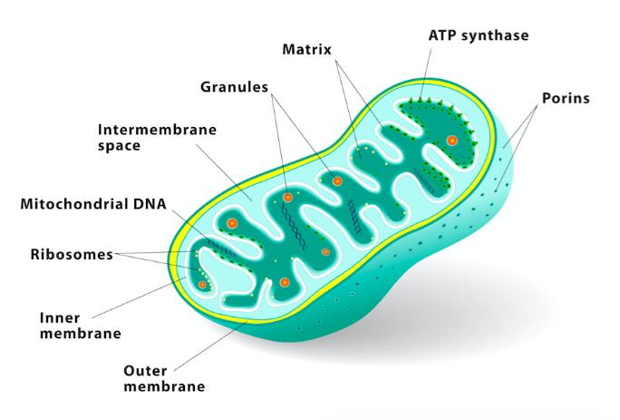
Extensive research has indicated that intracellular organelles are integrated into cellular networks and collaborate on various cellular tasks rather than acting as isolated entities. Specialized membrane contact sites are formed between organelles, providing distinct spatial regions for executing and regulating their functions. Mitochondria is crucial dynamic organelles that participate extensively in many important cellular processes. Its dysfunction has been implicated in diverse diseases such as neurodegenerative disorders, cancer, and cardiovascular diseases.


Capturing the dynamic process of subcellular organelles in living cells is crucial to the study of disease mechanism. The limitations of existing fluorescent mitochondrial probes, such as photobleaching and a nonspecific background, hinder the characterization of dynamic physical interactions between mitochondria and other subcellular organelles in live cells.


Based on this research pain point, Abbkine innovatively developed TraKine™ Pro mitochondrial fluorescence probe-TraKine™ Pro Live-cell Mitochondrion Staining kit (Deep Red Fluorescence with Super Resolution)


Product nameCat#Ex/Em(nm)Live or fixed cellsMicroscope compatibility
TraKine™ Pro Live-cell Mitochondrion Staining kit (Deep Red Fluorescence with Super Resolution)KTC4300647/661Live& fixed cellsFluorescence microscope,Confocal, STED, SIM, PALM, STORM, TIRF


【Advantages】

 
  • Optimized lysosomal staining scheme for living and fixed cells of mammals

  • It is especially suitable for living cell research under laser Confocal microscope and long-term super-resolution microscope (such as SIM, STED, TIRF, STORM, PALM), and is perfectly used for observing cell dynamics in three-dimensional space.

  • Patented dye MitoRed™ (Ex/EM=647/661 nm)-High specificity, low background, perfect fluorescence stability

  • Low levels of cytotoxicity


【Results】


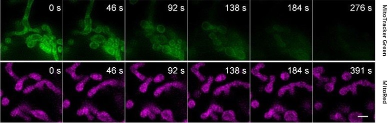
一: Compared with commercially available MitoTracker® Green (ThermoFisher), TraKine™ Pro Live-cell Mitochondrion Fluorescence Probe shows better light resistance and lower bleaching property.


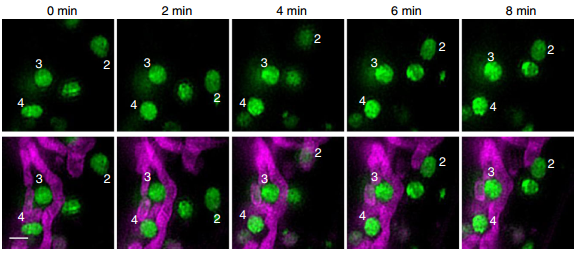
二: SIM images of lysosomes and mitochondria of U2OS living cells using TraKine™ Pro Live-cell Lysosome Staining kit (Green Fluorescence with Super Resolution) and TraKine™ Pro Live-cell Mitochondrion Staining kit (Deep Red Fluorescence with Super Resolution) reveal the dynamic interaction process of lysosomes and mitochondria.


About Abbkine Scientific Co., Ltd.

Abbkine serves global scientists in the field of proteomics and cytology and is committed to the innovation and development of various scientific reagents related to proteomics and cytology, expecting to accelerate the pace of life science research and drug discovery. Proteomics products cover the preparation of samples (protein extraction, purification, coupling), protein quantification, antibodies and kits for protein detection. Cytology products involve cytokines (cell culture), cell status detection, cell staining, organelle extraction, cell metabolism and cytopathology reagents (kits). Abbkine relies on the product portfolio and unique marketing support as the main market strategy and product innovation mode, with ultimate aim to facilitate your research career.

2019年3月22日星期五

TraKine™ Cell Staining Kits

Full portfolio for staining of cell and organelles including Tubulin, Mitochondrion, Lysosome, Nuclei, Plasma Membrane and so on.


[pdfjs-viewer url="https%3A%2F%2Fwww.abbkine.com%2Ffile%2Fdistributor%2FTraKine%20cell%20staining.pdf" viewer_width=100% viewer_height=1360px fullscreen=true download=false print=true]

Abbkine Top Selling Products for Proteomics

Abbkine focuses on Proteomics and Cytology. Here, we present our top selling products in proteomics research, from basic products such as protein extraction, protein purification, protein coupling, protein quantification, antibodies and kits for protein detection, to help your research career. The following products are available in stock.

[pdfjs-viewer url="https%3A%2F%2Fwww.abbkine.com%2Ffile%2Fdistributor%2FAbbkine%20Top%20Selling%20Products%20for%20Proteomics.pdf" viewer_width=100% viewer_height=1360px fullscreen=true download=false print=true]

2019年3月15日星期五

Living cell staining, super-resolution Lysosome tracing

【REVIEW】:


lysosome

Lysosome is an important organelle in animal cells. Its integrity is closely related to animal physiology and pathology. Lysosomes are vesicle-like organelles surrounded by a single membrane and containing a variety of acidic hydrolases. Their main function is to carry out intracellular digestion and specialize in decomposing various exogenous and endogenous macromolecular substances. Lysosomes in cells are heterogeneous, and the size and types of hydrolytic enzymes contained in lysosomes may vary greatly. The marker enzyme is acid phosphatase. Recent studies have found that phagocytes and lysosomes have bidirectional signaling functions. The ability of lysosomes to move towards transport vesicles is crucial for the degradation of biological macromolecules.


Unfortunately, the lysosomal dyes commonly used in the market are only suitable for the observation of cells on the plane, rather than the three-dimensional structure of cells. You will always see only uniform but featureless clouds of colors. A specific dye compatible with long-time super-resolution microscopic imaging technology (such as SIM, STED, TIRF, STORM, PALM) has become a research reagent urgently needed by researchers.


Abbkine has innovatively developed TraKine™ Pro series lysosome-specific fluorescent staining kit, in response to this research difficulty.


Product nameCat#Microscope compatibility
TraKine™ Pro Live-cell Lysosome Staining kit (Green Fluorescence with Super Resolution)KTC4200Fluorescence microscope,Confocal, STED, SIM, PALM, STORM, TIRF
TraKine™ Pro Live-cell Lysosome Staining kit (Deep Red Fluorescence with Super Resolution)KTC4210Fluorescence microscope,Confocal, SIM, PALM, STORM, TIRF


Confocal (confocal microscopes

STED(stimulated emission depletion)

SIM(structured-illumination microscopy)

PALM(photoactivation localization microscopy)

STORM(stochastic optical reconstruction microscopy)

TIRF(total internal reflection fluorescence imaging)


Take TraKine™ Pro Live-cell Lysosome Staining kit (Green Fluorescence with Super Resolution) for example:


Principle

TraKine™ Pro Live-cell Lysosome Staining kit (Green Fluorescence with Super Resolution) is a treasure-grade fluorescence imaging tool for staining lysosomes in living and fixed cells of mammals, with high specificity and low background. The patented probe in the kit consists of lysosome recognition unit and green fluorescent dye (Ex/Em = 494/517nm), and has high fluorescence quantum yield and high light stability. Probes can selectively recognize and label lysosomes. It is especially suitable for long-term super-resolution imaging.


【Advantage】
  • Optimized lysosomal staining scheme for living and fixed cells of mammals.

  • It is especially suitable for living cell research under laser Confocal microscope and long-term super-resolution microscope (such as SIM, STED, TIRF, STORM, PALM), and is perfectly used for observing cell dynamics in three-dimensional space.

  • Patented dye LysGreen™ (Ex/EM=494/517 nm)-High specificity, low background, perfect fluorescence stability.

  • Almost no cytotoxicity.


【Result】


SIM imaging: U2OS cells, greenConfocal imaging: U2OS cells, green


U2OS cells, dynamic SIM imaging results (Red)U2OS cells, dynamic SIM imaging results (Red)


*TraKine™ Pro is series of long-term super-resolution cell staining imaging portfolio for labeling subcellular structures of live and fixed cells. TraKine™ Pro proprietary excellent fluorescent dyes span the full UV-visible and near IR spectrum.


About Abbkine Scientific Co., Ltd.


Abbkine serves global scientists in the field of proteomics and cytology and is committed to the innovation and development of various scientific reagents related to proteomics and cytology, expecting to accelerate the pace of life science research and drug discovery. Proteomics products cover the preparation of samples (protein extraction, purification, coupling), protein quantification, antibodies and kits for protein detection. Cytology products involve cytokines (cell culture), cell status detection, cell staining, organelle extraction, cell metabolism and cytopathology reagents (kits). Abbkine relies on the product portfolio and unique marketing support as the main market strategy and product innovation mode, with ultimate aim to facilitate your research career.

2019年3月11日星期一

TraKine™ Pro Live-cell Tubulin Staining kit-Your Best Choice for Cytoskeleton Study

【Review 】

TraKine™ Pro Live-cell Fluorescence Tool-Perfect compatibility with long-term super-resolution fluorescence imaging

As soon as the Abbkine TraKine™ Pro fluorescent dyes were released, they received high attention from living cell imaging researchers all over the world. With extremely high specificity, the probes can last for several hours in living cells, which is particularly suitable for studying the dynamics and interactions of subcellular structures in live cells for super-resolution imaging.

In eukaryotes, microtubules are one of the main components of cytoskeleton. Cytoskeleton is not only involved in supporting cells, but also related to almost all forms of cell movements, such as cell migration, transport of organelles and macromolecules. In addition, it is closely related to signal transduction and cell division. Tubulin is the basic structural unit of intracellular microtubules. There are two types of Tubulin, α-tubulin and β-tubulin. The two Tubulin proteins have similar three-dimensional structure and can be tightly combined into dimers to serve as subunits of microtubule assembly.


Cytoskeleton is a dynamic structure and plays an important role in many life processes. Immunofluorescence staining is very common in cytoskeletal studies, but it is mainly applicable to fixed or transmembrane treated cells, and cannot track the dynamic activities of living cells very well. Fusion fluorescent protein is also a labeling method, but it has been reported that fusion fluorescent protein has side effects on cell extension, migration and adhesion.

Abbkine TraKineTM Pro Live-cell Tubulin Staining kit is a fluorescence imaging tool for staining of Tubulin in mammalian living cells with high specificity and low background. The proprietary probe in the kit consists of a microtubule recognition unit and a fluorescent dye, the recognition unit can selectively recognize microtubules and binding with it. The whole probe is membrane impermeable, but after incubating with buffer T, it can enter cells through endocytosis pathway in vesicular compartments, and release into cytoplasm subsequently. It is especially suitable for Confocal and long-term super-resolution imaging (such as SIM, STED, TIRF, STORM and PALM).


Product nameCAT #:Ex/Em(nm)
TraKine™ Pro Live-cell Tubulin Staining kit
(Green Fluorescence with Super Resolution)
KTC4100500/520
TraKine™ Pro Live-cell Tubulin Staining kit (Deep Red Fluorescence with Super Resolution)KTC4110650/665


Features & Benefits

Living cells labeling: Optimized staining protocol for labeling Tubulin in mammalian living cells

Stable and persistent: fluorescence can last for several hours and is ideal for monitoring dynamic

Safe: Low levels of cytotoxicity to cells

Super- resolution:especially suitable for Confocal and long-term super-resolution imaging (such as SIM, TIRF, STORM and PALM)

Verification system:has been tested in U2OS, APRE, Hela, 3T3 and cos-7 cell lines

Customizable: span the full UV-visible and near IR spectrum,

Perfect results are the best illustration


SIM imaging results of U2OS cells using Abbkine TraKine™ Pro Live-cell Tubulin Staining kit. The filamentous structure of microtubules can be clearly labeled.

2019年3月8日星期五

TraKine™ Pro Live-cell Fluorescence Tool-Perfect compatibility with long-term super-resolution fluorescence imaging

Combining genetic marker method with small and bright probes that can be controlled by light-controlled switches will be a very ideal method to develop molecular-level ultra-high resolution imaging in the future.

- Xiaowei Zhuang (Discoveries of novel cellular structures using STORM)

Inspired by academician Xiaowei Zhuang, Abbkine TraKine™ Pro series of fluorescent dyes came into being.
TraKine™ Pro is series of long-term super-resolution cell staining imaging portfolio for labeling subcellular structures of live and fixed cells. TraKine™ Pro proprietary excellent fluorescent dyes span the full UV-visible and near IR spectrum.

This release of the TraKine™ Pro series featured hot research targets, including,

Tubulin: One of the important components of eukaryotic cytoskeleton, plays an important role in maintaining cell morphology, cell division, signal transduction and material transport.
Lysosome & Mitochondrion: An important dynamic organelle, widely involved in many important cellular processes. Their dysfunction is related to many diseases, such as neurodegenerative diseases, cancer and cardiovascular diseases.
Nuclei: Controls the genetic characteristics of organisms. Cell metabolism is mainly carried out by controlling the synthesis of specific enzymes. Responsible for protein synthesis, cell division, growth and differentiation, and regulating gene integrity and gene expression.

* We accept product customization based on TraKine™ Pro technology.

 Preemptive attention to product parameters:


Product nameCat#TargetEx/Em(nm)Live or fixed cellsMicroscope compatibility
TraKine™ Pro Live-cell Tubulin Staining kit (Green Fluorescence with Super Resolution)KTC4100Tubulin500/520Live cellsFluorescence microscope,Confocal, STED, SIM, PALM, STORM, TIRF
TraKine™ Pro Live-cell Tubulin Staining kit (Deep Red Fluorescence with Super Resolution)KTC4110Tubulin650/665Live cellsFluorescence microscope,Confocal, SIM, PALM, STORM, TIRF
TraKine™ Pro Live-cell Lysosome Staining kit (Green Fluorescence with Super Resolution)KTC4200Lysosome494/517Live& fixed cellsFluorescence microscope,Confocal, STED, SIM, PALM, STORM, TIRF
TraKine™ Pro Live-cell Lysosome Staining kit (Deep Red Fluorescence with Super Resolution)KTC4210Lysosome650/665Live& fixed cellsFluorescence microscope,Confocal, SIM, PALM, STORM, TIRF
TraKine™ Pro Live-cell Mitochondrion Staining kit (Deep Red Fluorescence with Super Resolution)KTC4300Mitochondrion647/661Live& fixed cellsFluorescence microscope,Confocal, STED, SIM, PALM, STORM, TIRF
TraKine™ Pro Live-cell Nuclei Staining kit (Green Fluorescence with Super Resolution)KTC4500Nuclei494/517Live& fixed cellsFluorescence microscope,Confocal, STED, SIM, PALM, STORM, TIRF
TraKine™ Pro Live-cell Nuclei Staining kit (Deep Red Fluorescence with Super Resolution)KTC4510Nuclei650/665Live& fixed cellsFluorescence microscope,Confocal, SIM, PALM, STORM, TIRF

Confocal: confocal microscopes

STED: stimulated emission depletion

SIM: structured-illumination microscopy

PALM: photoactivation localization microscopy

STORM: stochastic optical reconstruction microscopy

TIRF,: total internal reflection fluorescence imaging

If you only limit the observation of cells on the plane instead of the three-dimensional structure of cells, you will always see only consistent but featureless clouds of color. Using unique TraKine™ Pro series dyes, combined with super-resolution microscopy technology, the real cell structure and fresh and living colors spring up!

The choice of beauty or mediocrity depends entirely on the horizon. Feel the unique charm of TraKine™ Pro series dyes together.


SIM imaging results of U2OS cells using Abbkine TraKine™ Pro Live-cell Tubulin Staining kit (Green Fluorescence with Super Resolution). It can be seen that the kit can specifically label microtubule structures in living cells and perform ultra-high resolution imaging.


SIM imaging results of U2OS cells using Abbkine TraKine™ Pro Live-cell Lysosome Staining kit (Deep Red Fluorescence with Super Resolution). It can be seen that the kit can specifically label Lysosome structures in living cells and perform ultra-high resolution imaging.


SIM imaging results of U2OS cells using Abbkine TraKine™ Pro Live-cell Mitochondrion Staining kit (Deep Red Fluorescence with Super Resolution).


About Abbkine Scientific Co., Ltd.

 Abbkine serves global scientists in the field of proteomics and cytology and is committed to the innovation and development of various scientific reagents related to proteomics and cytology, expecting to accelerate the pace of life science research and drug discovery. Proteomics products cover the preparation of samples (protein extraction, purification, coupling), protein quantification, antibodies and kits for protein detection. Cytology products involve cytokines (cell culture), cell status detection, cell staining, organelle extraction, cell metabolism and cytopathology reagents (kits). Abbkine relies on the product portfolio and unique marketing support as the main market strategy and product innovation mode, with ultimate aim to facilitate your research career.