2019年9月24日星期二

In 4 years, Abbkine helped more than 1200 English articles to be published. Impact factors exceed 4500 points! ! !

Abbkine focuses on immunology and cytology, and is committed to innovating and developing various antibodies, proteins, analytical reagents and kits, with a view to becoming a key promoter in the fields of life science research and development, drug research and development, etc.


Some articles published using Abbkine products:


1. The negative effect of silica nanoparticles on adipogenic differentiation of human mesenchymal stem cells. Materials Science and Engineering. (IF: 27.24)


Using Abbkine Dylight 594, Goat Anti-Mouse IgG


2. Bph6 encodes an exocyst-localized protein and confers broad resistance to planthoppers in rice. NATURE GENETICS. (IF: 19.88)


Using Abbkine Anti-Plant Actin Mouse Monoclonal Antibody (3T3)


3. The deubiquitinating enzyme cylindromatosis mitigates nonalcoholic steatohepatitis. . (IF: 19.14)


Using Abbkine HRP, Goat Anti-Rabbit IgG


4. Research on the interaction of protein Tmub1 and securin after partial hepatectomy. Journal of the American Chemical Society. (IF: 14.75)


Using Abbkine IFKine™ Red Donkey Anti-Rabbit IgG


5. Verification of Differential Expression Genes after CacyBP/SIP Nuclear Translocation in Colon Carcinoma Cell Line. Journal of the American Chemical Society. (IF: 14.75)


Using Abbkine Anti-PCNA Mouse Monoclonal Antibody (1D7)


6. CD8+T cells induce cachexia during chronic viral infection. NATURE IMMUNOLOGY. (IF: 14.71)


Using Abbkine EliKine™ Free Triiodothyronine (fT3) ELISA Kit


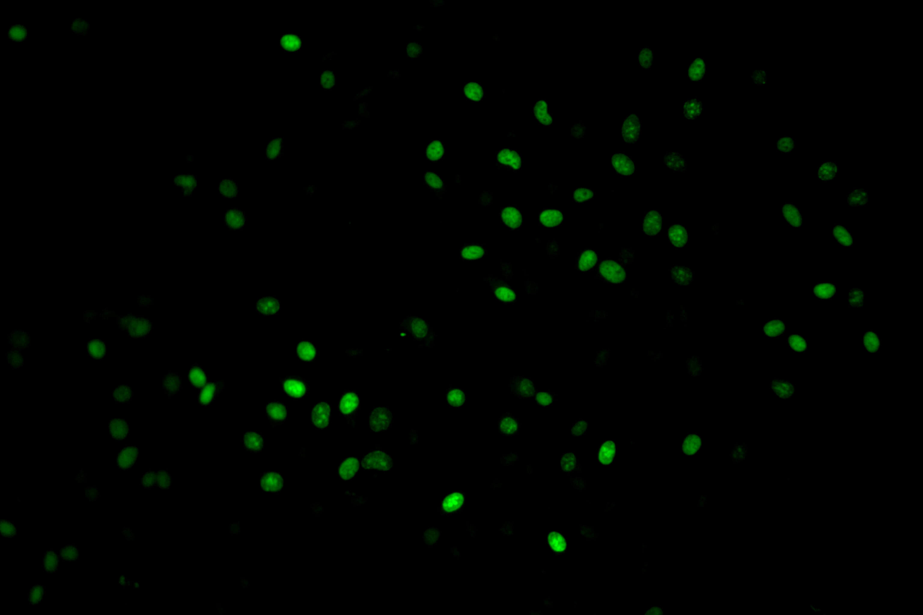
7. SET8 prevents excessive DNA methylation by methylation-mediated degradation of UHRF1 and DNMT1. Nucleic Acids Research. (IF: 11.14)


Using Abbkine Pan Methylated Lysine Monoclonal Antibody (Mix)


8. The lncRNA PVT1 regulates nasopharyngeal carcinoma cell proliferation via activating the KAT2A acetyltransferase and stabilizing HIF-1α. Cell Death & Differentiation. (IF: 8.086)


Using Abbkine KAT6A
Polyclonal Antibody


9. DIPK2A promotes STX17- and VAMP7-mediated autophagosome-lysosome fusion by binding to VAMP7B. Autophagy. (IF: 7.01)


Using Abbkine IPKine™
HRP, Goat Anti-Mouse IgG HCS


10. CAV1-CAVIN1-LC3B-mediated autophagy regulates high glucose-stimulated LDL transcytosis. Autophagy. (IF: 7.01)


Using Abbkine IPKine™ HRP, Goat Anti-Rabbit IgG HCS


Some partners of
Abbkine:


Harvard University, Georgia Institute of Technology, Oxford University, Duke University, Stanford University, Peking University, Tsinghua University, Zhejiang University, Chinese Academy of Sciences, Wuhan University, Fudan University.


2019年9月10日星期二

Research Hotspots of Cell Apoptosis: Bcl-2 Monoclonal Antibody

【Promotion】Abbkine
Selected Primary Antibody-Click for Promotion
details.


Bcl-2 gene (B-cell lymphoma-2) is a member of Bcl-2 family, which is the earliest discovered apoptosis protein. It is an anti-apoptosis protein containing multiple BH domains. It is highly expressed in some cancer cells and selectively plays an anti-tumor role. Bcl-2 family proteins play an important role in the regulation of apoptosis by forming dimers with Bax and dimerizing themselves. When Bcl-2 protein is inhibited, its dimer with Bax decreases, leading to cell apoptosis. When Bcl-2 protein is overexpressed, heterodimers formed by Bcl-2 protein and Bax increase and apoptosis is inhibited. The balance between Bcl-2 and Bax proteins at the cell death signal checkpoint determines the survival or apoptosis of cells.


Fig: Bcl-2 and Cell apoptosis

We recommend Abbkine selected Bcl-2 antibody for WB, IHC-P and IF experiments in human, rats, mice and chickens.


Product name

Cat#

Application

MW

Bcl-2 Monoclonal Antibody

ABM0010

WB(1:1000-2000),IF(1:50-200),IHC-P(1:200)

26KD


The result:


1) Hela cells; 2) MCF-7 cells; Using Abbkine Bcl-2 monoclonal antibody (ABM0010), WB 1:1000 dilution.


Human oviduct, Using Abbkine Bcl-2 monoclonal antibody (ABM0010) and IHC-P 1:100.


NIH/3T3 cells, Using Abbkine Bcl-2 monoclonal antibody (ABM0010), green, IF 1:100.

2019年9月2日星期一

Beyond BrdU, New Generation Cell Proliferation Imaging Analysis Kit (EdU Method)

Common methods of cell proliferation
mainly include MTT method, WST-1 method and CCK-8 method, which are mainly
based on the activity of cells, thus reflecting the overall proliferation
effect, but unable to detect individual proliferating cells. Recognized as the
most accurate method to detect cell proliferation is to directly detect DNA
synthesis in cells. Previously, BrdU method was the most commonly used method.
However, the method requires DNA denaturation (such as acid denaturation,
thermal denaturation or DNase digestion) to expose BrdU, thus binding to BrdU
antibody. The whole experiment has many influencing factors and poor stability.


Abbkine innovates and develops EdU
method based on EdU incorporation and subsequent click reaction to solve this
application difficulty:


Product name

Cat#

Fluorescent characteristic

Cell Proliferation EdU Image Kit
(Green Fluorescence)

KTA2030

AbFluor
488
Ex/Em = 501/525 nm

Cell Proliferation EdU Image Kit
(Orange Fluorescence)

KTA2031

AbFluor
545
Ex/Em = 546/565 nm


Principle:EdU (5-ethynyl-2´-deoxyuridine) is a nucleoside analog of thymidine and is incorporated into DNA during active DNA synthesis. Comparing to BrdU assays, the EdU-Click Assays are not antibody based and therefore do not require DNA denaturation (typically using HCl or heat or digestion with DNase) for detection of the incorporated nucleoside. Detection is based on a click reaction, a copper-catalyzed covalent reaction between an azide and an alkyne, is complete within 30 minutes.


Kit components

Advantage
• EdU (10mM)
• AbFluor 488 azide or AbFluor 545 azide
• 10×Reaction buffer
• Copper
• Reducing Agent
• Not antibody based 
• No DNA denaturation. Can maintain cell morphology and DNA integrity
• Patented AbFluor 488 and 545 azide have good light stability and quenching resistance
• Optimization for fluorescence microscope


Results:


KTA2030
Imaging: Hela Cells (Green)

KTA2031
Imaging: Hela Cells (Red)