2019年11月4日星期一

Abbkine winter promotion: looking for you who need live-cell probe

Promotion time: From now to 15th January, 2020.


Promotion product
list:
TraKine™ Pro product
line (Tubulin, Lysosome, Mitochondrion, Nuclei live-cell probe ).


Details of activities: 20% off for TraKine™ Pro product line.


Details for promotion product:


Product name

#Cat

Target

Ex/Em (nm)

Size

Promotion price ($)

TraKine™ Pro Live-cell Tubulin Staining
kit (Green Fluorescence with Super Resolution)

KTC4100

Tubulin

500/520

50/250T

144/496

TraKine™ Pro Live-cell Lysosome Staining
kit (Deep Red Fluorescence with Super Resolution)

KTC4210

Lysosome

650/665

50/250T

128/472

TraKine™ Pro Live-cell Lysosome Staining
kit (Orange Fluorescence with Super Resolution)

KTC4220

Lysosome

565/590

50/250T

128/4721

TraKine™ Pro Live-cell Mitochondrion
Staining kit (Deep Red Fluorescence with Super Resolution)

KTC4300

Mitochondrion

647/661

50/250T

176/576

TraKine™ Pro Live-cell Nuclei Staining
kit (Deep Red Fluorescence with Super Resolution)

KTC4510

Nuclei

650/665

50/250T

144/496


TraKine™ Pro features & benefits:


  • Proprietary probe contains fluorescent dye and a unit which
    electively recognize organelles, with high specificity, low background,
    excellent photostability and good cell permeability.
  • Optimized staining protocol for labeling subcellular structures in
    mammalian living and fixed cells.
  • Fuorescence can last for several hours in cells, stable and
    persistent, especially ideal for monitoring dynamics.
  • Super resolution, especially suitable for Confocal and long-term
    super-resolution imaging (such as SIM, STED, TIRF, STORM and PALM).
  • Safe, very low or no cytotoxicity to cells.

2019年10月30日星期三

Abbkine P53 antibody helps basic cancer research!

P53 gene, human tumor suppressor gene. The gene encodes a protein with a molecular weight of 43.7KDa, but is named P53 because the protein band appears at 53KDa shown by Marker. Wild-type p53 causes cancer cells to apoptosis, thus preventing canceration; But also has the function of helping cell genes repair defects; Studies have found that mutant p53 can enhance canceration. 50% of human tumors are related to P53 gene. Currently, there are liver cancer, breast cancer, bladder cancer, gastric cancer, colon cancer, prostate cancer, soft tissue sarcoma, ovarian cancer, brain tumor, lymphocyte tumor, esophageal cancer, lung cancer, osteosarcoma, etc. P53 mutation in human tumors is mainly in highly conserved regions, with the highest mutation at 175, 248, 249, 273, 282 sites, and different types of tumors.  


P53 has a very short half-life, so it is almost undetectable in unstressed cells. This is mainly realized by the continuous ubiquitination of p53 and the degradation of 26S proteasome in the later stage under the action of its inhibitor MDM2. Cell stress signals such as DNA damage can inhibit ubiquitination of p53 and lead to its stabilization and activation. Once p53 is activated, it will bind to its downstream target regulatory elements and regulate its transcription. Therefore, various cell therapy schemes based on different tumor inhibition functions of p53 can be started. Just because past studies have shown that p53 gene mutations exist in about half of cancers, scientists regard them as important targets for developing new cancer therapies.


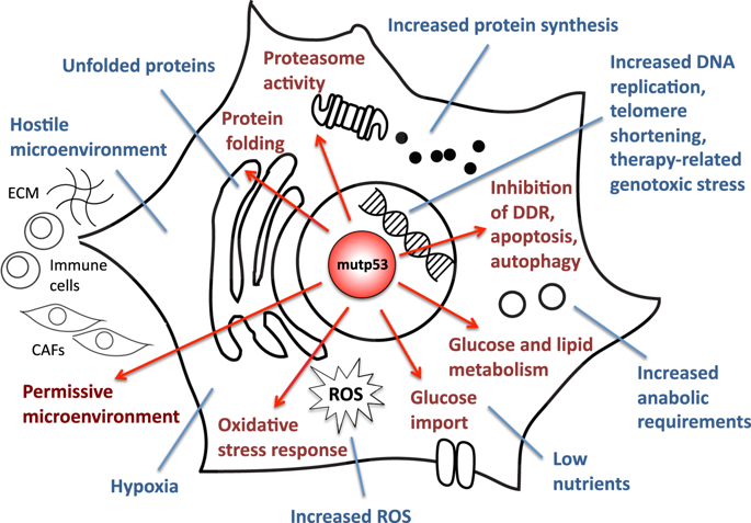
Mutant p53 promotes adaptive responses to cancer-related stress conditions to support tumor progression.

Abbkine P53 Antibody Helps Basic Cancer
Research! Basic information about the product:


Product name
#CAT
Reactivity Applications
Dilution ratio
p53 Polyclonal Antibody ABP0110   Human, Monkey, Mouse, Rat
WB/IF/IHC-P/ELISA

WB (1:500-1:2000)
IF (1:200-1:1000)
IHC-P (1:100-1:300)
ELISA  (1:10000).

Abbkine P53 Antibody verification results:


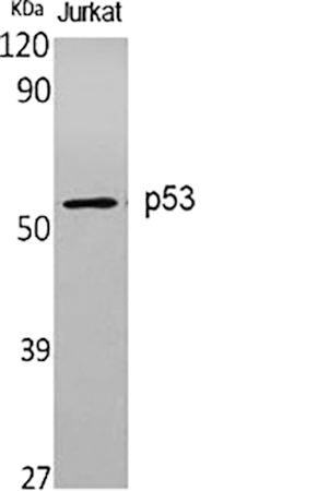
Western Blot analysis of various cells using p53 Polyclonal Antibody diluted at 1:1000.
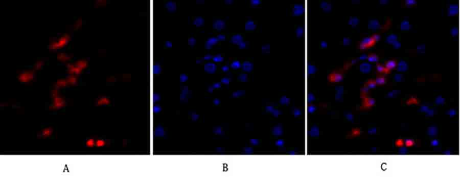
Immunofluorescence analysis of human liver tissue. 1, p53 Polyclonal Antibody (red) was diluted at 1:200 (4°C, overnight). 2, Cy3 Labeled secondary antibody was diluted at 1:300 (room temperature, 50min). 3, Picture B: DAPI (blue) 10min. Picture A: Target. Picture B: DAPI. Picture C: merge of A+B.
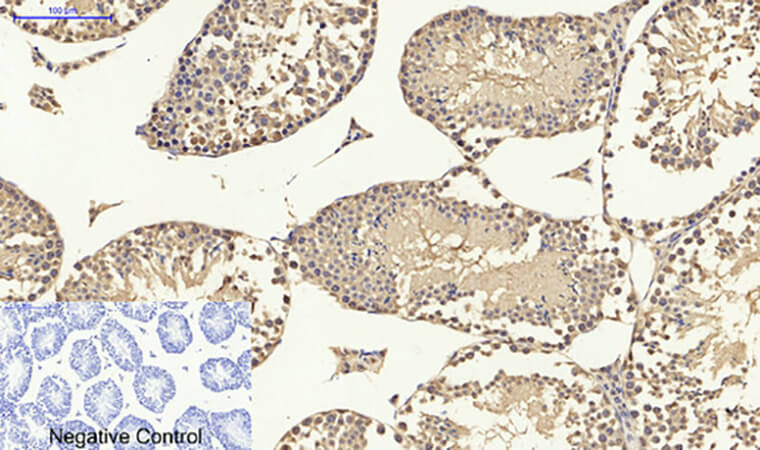
Immunohistochemical analysis of paraffin-embedded mouse testis tissue. 1, p53 Polyclonal Antibody was diluted at 1:200 (4°C, overnight). 2, Sodium citrate pH 6.0 was used for antibody retrieval (>98°C, 20min). 3, secondary antibody was diluted at 1:200 (room temperature, 30min). Negative control was used by secondary antibody only.

2019年10月15日星期二

IL-6: Target Molecule for Cytokine Storms

Interleukin-6 (IL-6) is a multi-effect cytokine
that plays an important role in host defense by regulating immune and
inflammatory reactions. IL-6 is mainly produced by macrophages, T lymphocytes
and B lymphocytes. When the body has inflammatory reaction or infection,
various cytokines such as virus, endotoxin and tumor necrosis factor can induce
the body to produce IL-6. The biological effects of IL-6 are mainly reflected
in inducing B cell differentiation to produce immunoglobulin, promoting T cell
proliferation and growth, enhancing blood cell differentiation and its
anti-tumor effect, promoting bone marrow hematopoietic stem cell proliferation,
etc.


IL-6 is an important member of cytokine network and plays a central role in acute inflammatory reaction. The production of IL-6 induces the production of C-reactive protein (CRP) and procalcitonin (PCT), which are directly related to inflammatory diseases and infection degree. IL-6 can diagnose early inflammation and warn the occurrence of sepsis faster. In addition, the half-life of IL-6 is shorter than CRP and PCT, which can reflect the effect of antibiotic treatment faster and better reflect the prognosis of patients. At the same time, IL-6 can be used as a biomarker of disease severity and prognosis indicators in cytokine storms, and its expression is superior to TNF-α and IL-1. A large number of research results show that IL-6 is a suitable target molecule for cytokine storms.


If you are studying IL-6 or are about to study IL-6, the recommended IL-6 cytokines are as follows:


Product
name


#cat

Sequence

Purity

Expressed
Host


Endotoxin

Formulation

Human
IL-6 protein

PRP100120

Human IL6
(NP_000591.1) (Val 30-Met 212)

>
95%

E.
coli

<1EU/μg

lyophilized
powder

Mouse IL-6 protein

PRP100371

Mouse
IL6 (P08505) (Phe25-Thr211)

>
95%

E.
coli

<1EU/μg

lyophilized
powder

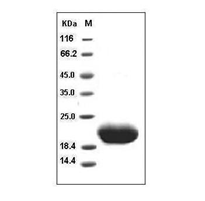
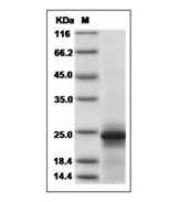
SDS-PAGE analysis:


Human IL-6 protein


Mouse IL-6 protein


Recommendations
for cytokine storage:


Cytokines
in lyophilized powder form are very stable and can be stored for several years
at -20℃ or -80℃. Freeze-dried powder should be dissolved before use and then
added to the culture medium in liquid form. The dissolved cytokines can only be
stored for a short time (about 1 week) at 4℃. For long-term preservation, first
prepare a diluent (which must contain carrier protein, such as 0.1% BSA, 5%HSA,
or 10% FBS), and then sub-pack and freeze at -20℃ or -80℃. Repeated freezing
and thawing must be avoided, as each freezing and thawing will cause partial
inactivation of proteins.

2019年10月10日星期四

Abbkine Meets You at 17th International Immunology Congress (IUIS 2019)

Abbkine
invites you to visit IUIS 2019, Beijing, China.


Abbkine featured products and services will be displayed at the exhibition. There always be one type can flash into your eyes. Participate in interaction, more surprises are waiting for you!


Date: October
19 – 23, 2019


Booth:#D22,level 4


Address: China
National Convention Center, Beijing, China


About: The 17th International Congress of Immunology (IUIS 2019) will be held from October 19 – 23, 2019 at the China National Convention Center, Beijing, China. IUIS 2019 promises to be an unforgettable event that will bring together delegates from all over the world. We anticipate over 5000 participants, including international leaders at the forefront of the discipline that will present the most recent advances in basic immunology and clinical translation.


2019年9月24日星期二

In 4 years, Abbkine helped more than 1200 English articles to be published. Impact factors exceed 4500 points! ! !

Abbkine focuses on immunology and cytology, and is committed to innovating and developing various antibodies, proteins, analytical reagents and kits, with a view to becoming a key promoter in the fields of life science research and development, drug research and development, etc.


Some articles published using Abbkine products:


1. The negative effect of silica nanoparticles on adipogenic differentiation of human mesenchymal stem cells. Materials Science and Engineering. (IF: 27.24)


Using Abbkine Dylight 594, Goat Anti-Mouse IgG


2. Bph6 encodes an exocyst-localized protein and confers broad resistance to planthoppers in rice. NATURE GENETICS. (IF: 19.88)


Using Abbkine Anti-Plant Actin Mouse Monoclonal Antibody (3T3)


3. The deubiquitinating enzyme cylindromatosis mitigates nonalcoholic steatohepatitis. . (IF: 19.14)


Using Abbkine HRP, Goat Anti-Rabbit IgG


4. Research on the interaction of protein Tmub1 and securin after partial hepatectomy. Journal of the American Chemical Society. (IF: 14.75)


Using Abbkine IFKine™ Red Donkey Anti-Rabbit IgG


5. Verification of Differential Expression Genes after CacyBP/SIP Nuclear Translocation in Colon Carcinoma Cell Line. Journal of the American Chemical Society. (IF: 14.75)


Using Abbkine Anti-PCNA Mouse Monoclonal Antibody (1D7)


6. CD8+T cells induce cachexia during chronic viral infection. NATURE IMMUNOLOGY. (IF: 14.71)


Using Abbkine EliKine™ Free Triiodothyronine (fT3) ELISA Kit


7. SET8 prevents excessive DNA methylation by methylation-mediated degradation of UHRF1 and DNMT1. Nucleic Acids Research. (IF: 11.14)


Using Abbkine Pan Methylated Lysine Monoclonal Antibody (Mix)


8. The lncRNA PVT1 regulates nasopharyngeal carcinoma cell proliferation via activating the KAT2A acetyltransferase and stabilizing HIF-1α. Cell Death & Differentiation. (IF: 8.086)


Using Abbkine KAT6A
Polyclonal Antibody


9. DIPK2A promotes STX17- and VAMP7-mediated autophagosome-lysosome fusion by binding to VAMP7B. Autophagy. (IF: 7.01)


Using Abbkine IPKine™
HRP, Goat Anti-Mouse IgG HCS


10. CAV1-CAVIN1-LC3B-mediated autophagy regulates high glucose-stimulated LDL transcytosis. Autophagy. (IF: 7.01)


Using Abbkine IPKine™ HRP, Goat Anti-Rabbit IgG HCS


Some partners of
Abbkine:


Harvard University, Georgia Institute of Technology, Oxford University, Duke University, Stanford University, Peking University, Tsinghua University, Zhejiang University, Chinese Academy of Sciences, Wuhan University, Fudan University.


2019年9月10日星期二

Research Hotspots of Cell Apoptosis: Bcl-2 Monoclonal Antibody

【Promotion】Abbkine
Selected Primary Antibody-Click for Promotion
details.


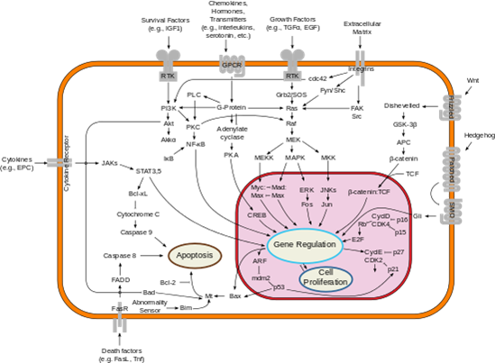
Bcl-2 gene (B-cell lymphoma-2) is a member of Bcl-2 family, which is the earliest discovered apoptosis protein. It is an anti-apoptosis protein containing multiple BH domains. It is highly expressed in some cancer cells and selectively plays an anti-tumor role. Bcl-2 family proteins play an important role in the regulation of apoptosis by forming dimers with Bax and dimerizing themselves. When Bcl-2 protein is inhibited, its dimer with Bax decreases, leading to cell apoptosis. When Bcl-2 protein is overexpressed, heterodimers formed by Bcl-2 protein and Bax increase and apoptosis is inhibited. The balance between Bcl-2 and Bax proteins at the cell death signal checkpoint determines the survival or apoptosis of cells.


Fig: Bcl-2 and Cell apoptosis

We recommend Abbkine selected Bcl-2 antibody for WB, IHC-P and IF experiments in human, rats, mice and chickens.


Product name

Cat#

Application

MW

Bcl-2 Monoclonal Antibody

ABM0010

WB(1:1000-2000),IF(1:50-200),IHC-P(1:200)

26KD


The result:


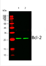
1) Hela cells; 2) MCF-7 cells; Using Abbkine Bcl-2 monoclonal antibody (ABM0010), WB 1:1000 dilution.


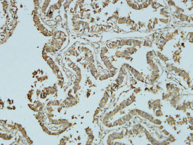
Human oviduct, Using Abbkine Bcl-2 monoclonal antibody (ABM0010) and IHC-P 1:100.


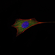
NIH/3T3 cells, Using Abbkine Bcl-2 monoclonal antibody (ABM0010), green, IF 1:100.

2019年9月2日星期一

Beyond BrdU, New Generation Cell Proliferation Imaging Analysis Kit (EdU Method)

Common methods of cell proliferation
mainly include MTT method, WST-1 method and CCK-8 method, which are mainly
based on the activity of cells, thus reflecting the overall proliferation
effect, but unable to detect individual proliferating cells. Recognized as the
most accurate method to detect cell proliferation is to directly detect DNA
synthesis in cells. Previously, BrdU method was the most commonly used method.
However, the method requires DNA denaturation (such as acid denaturation,
thermal denaturation or DNase digestion) to expose BrdU, thus binding to BrdU
antibody. The whole experiment has many influencing factors and poor stability.


Abbkine innovates and develops EdU
method based on EdU incorporation and subsequent click reaction to solve this
application difficulty:


Product name

Cat#

Fluorescent characteristic

Cell Proliferation EdU Image Kit
(Green Fluorescence)

KTA2030

AbFluor
488
Ex/Em = 501/525 nm

Cell Proliferation EdU Image Kit
(Orange Fluorescence)

KTA2031

AbFluor
545
Ex/Em = 546/565 nm


Principle:EdU (5-ethynyl-2´-deoxyuridine) is a nucleoside analog of thymidine and is incorporated into DNA during active DNA synthesis. Comparing to BrdU assays, the EdU-Click Assays are not antibody based and therefore do not require DNA denaturation (typically using HCl or heat or digestion with DNase) for detection of the incorporated nucleoside. Detection is based on a click reaction, a copper-catalyzed covalent reaction between an azide and an alkyne, is complete within 30 minutes.


Kit components

Advantage
• EdU (10mM)
• AbFluor 488 azide or AbFluor 545 azide
• 10×Reaction buffer
• Copper
• Reducing Agent
• Not antibody based 
• No DNA denaturation. Can maintain cell morphology and DNA integrity
• Patented AbFluor 488 and 545 azide have good light stability and quenching resistance
• Optimization for fluorescence microscope


Results:


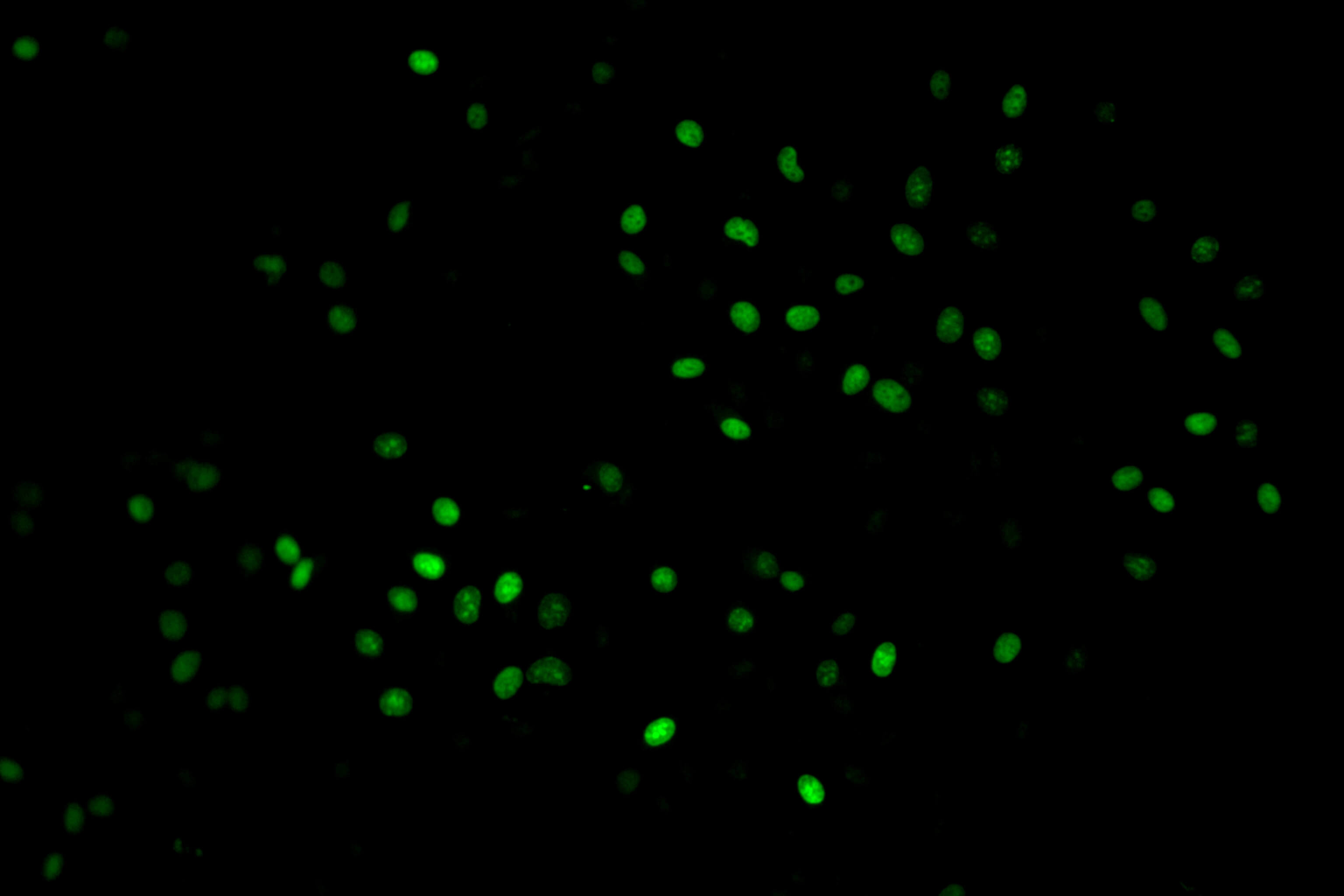
KTA2030
Imaging: Hela Cells (Green)

KTA2031
Imaging: Hela Cells (Red)