2019年11月27日星期三

Selected primary antibody|TNF-α antibody recommendation

Review of Related Articles:


Research
Hotspots of Cell Apoptosis: Bcl-2 Monoclonal Antibody


Abbkine
P53 antibody helps basic cancer research!


Marker of myofibroblast antibody-α-SMA/ACTA2 Monoclonal Antibody


Tumor necrosis factor (TNF, cachexin, or
cachectin; once named as tumor necrosis factor alpha or TNFα) is a cell
signaling protein (cytokine) involved in systemic inflammation and is one of
the cytokines that make up the acute phase reaction. It is produced chiefly by
activated macrophages, although it can be produced by many other cell types
such as CD4+ lymphocytes, NK cells, neutrophils, mast cells, eosinophils, and
neurons. TNF is a member of the TNF superfamily, consisting of various
transmembrane proteins with a homologous TNF domain.


The primary role of TNF is in the regulation of immune cells. TNF, being an endogenous pyrogen, is able to induce fever, apoptotic cell death, cachexia, inflammation and to inhibit tumorigenesis and viral replication and respond to sepsis via IL1- & IL6-producing cells. Dysregulation of TNF production has been implicated in a variety of human diseases including Alzheimer's disease, cancer, major depression, psoriasis and inflammatory bowel disease (IBD). Though controversial, studies of depression and IBD are currently being linked to increased levels of TNF. Recombinant TNF is used as an immunostimulant under the INN tasonermin. TNF can be produced ectopically in the setting of malignancy and parallels parathyroid hormone both in causing secondary hypercalcemia and in the cancers with which excessive production is associated.
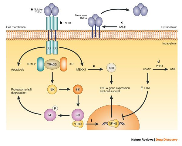


Fig: Anti-TNF-α therapies: the next generation (https://www.nature.com/articles/nrd1175)

Abbkine TNF-α
Polyclonal Antibody
helps basic cancer research! Basic information about
the product:


Product name

#CAT

Reactivity

Applications

Dilution ratio

Observed Band(KD)

TNF-α Polyclonal Antibody

ABP0127

Human/Mouse/Rat

WB/IF/IHC-P/ELISA

WB (1:500-1:2000)
IF (1:200-1:1000)
IHC-P (1:100-1:300)
ELISA(1/10000)

16KD

Abbkine TNF-α Polyclonal Antibody verification results:
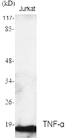


Western Blot analysis using Abbkine TNF-α Polyclonal Antibody.

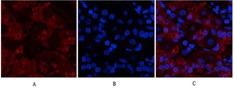
Immunofluorescence analysis of human
stomach tissue using Abbkine TNF-α Polyclonal Antibody (1:200).

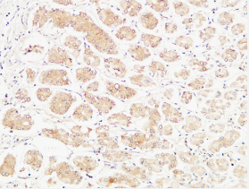
Immunohistochemical analysis of
paraffin-embedded Human stomach (1:100).

2019年11月14日星期四

Marker of myofibroblast antibody-α-SMA/ACTA2 Monoclonal Antibody

ACTA2 (actin alpha 2) is an actin protein
with several aliases including alpha-actin, alpha-actin-2, aortic smooth muscle
or alpha smooth muscle actin (α-SMA, SMactin, alpha-SM-actin, ASMA). Actins are
a family of globular multi-functional proteins that form microfilaments. ACTA2
is one of 6 different actin isoforms and is involved in the contractile
apparatus of smooth muscle. ACTA2 (as with all the actins) is extremely highly
conserved and found in nearly all mammals.


In humans, ACTA2 is encoded by the ACTA2
gene located on 10q22-q24. Mutations in this gene cause a variety of vascular
diseases, such as thoracic aortic disease, coronary artery disease, stroke,
Moyamoya disease, and multisystemic smooth muscle dysfunction syndrome.


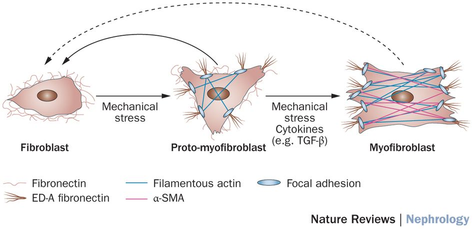
ACTA2 (commonly referred to as alpha-smooth muscle actin or α-SMA) is often used as a marker of myofibroblast formation.


Figure: Diverse origins of the
myofibroblast—implications for kidney fibrosis (From Nature Reviews)


Abbkine α-SMA Monoclonal Antibody Helps Basic Cancer Research! Basic information about the product:


Product name

#CAT

Reactivity

Applications

Dilution ratio

Observed Band(KD)

α-SMA Monoclonal
Antibody


ABM0052


Human/Mouse/ Rat

WB/IF/IHC-P

WB (1:10000-1:100000)
IF (1:100-1:200)
IHC-P (1:1000-1:2000)

42KD


Abbkine α-SMA Monoclonal Antibody verification results:


α-SMA-IF.png

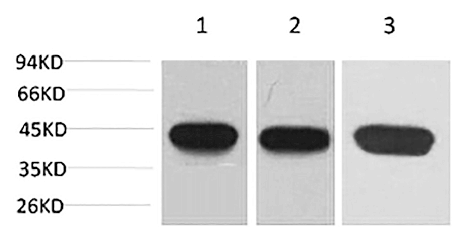
Western blot analysis of 1) Hela, 2) 3T3,
3) rat brain using α-SMA Monoclonal Antibody.
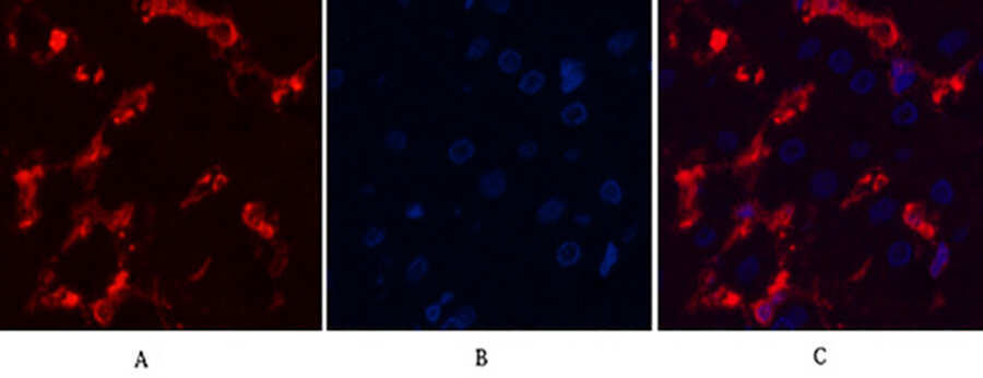
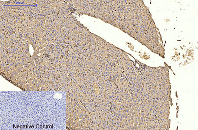
α-SMA-IHC

Immunofluorescence analysis of human
liver tissue.


α-SMA-WB

Immunohistochemical analysis of paraffin-embedded
mouse liver tissue

2019年11月4日星期一

Abbkine winter promotion: looking for you who need live-cell probe

Promotion time: From now to 15th January, 2020.


Promotion product
list:
TraKine™ Pro product
line (Tubulin, Lysosome, Mitochondrion, Nuclei live-cell probe ).


Details of activities: 20% off for TraKine™ Pro product line.


Details for promotion product:


Product name

#Cat

Target

Ex/Em (nm)

Size

Promotion price ($)

TraKine™ Pro Live-cell Tubulin Staining
kit (Green Fluorescence with Super Resolution)

KTC4100

Tubulin

500/520

50/250T

144/496

TraKine™ Pro Live-cell Lysosome Staining
kit (Deep Red Fluorescence with Super Resolution)

KTC4210

Lysosome

650/665

50/250T

128/472

TraKine™ Pro Live-cell Lysosome Staining
kit (Orange Fluorescence with Super Resolution)

KTC4220

Lysosome

565/590

50/250T

128/4721

TraKine™ Pro Live-cell Mitochondrion
Staining kit (Deep Red Fluorescence with Super Resolution)

KTC4300

Mitochondrion

647/661

50/250T

176/576

TraKine™ Pro Live-cell Nuclei Staining
kit (Deep Red Fluorescence with Super Resolution)

KTC4510

Nuclei

650/665

50/250T

144/496


TraKine™ Pro features & benefits:


  • Proprietary probe contains fluorescent dye and a unit which
    electively recognize organelles, with high specificity, low background,
    excellent photostability and good cell permeability.
  • Optimized staining protocol for labeling subcellular structures in
    mammalian living and fixed cells.
  • Fuorescence can last for several hours in cells, stable and
    persistent, especially ideal for monitoring dynamics.
  • Super resolution, especially suitable for Confocal and long-term
    super-resolution imaging (such as SIM, STED, TIRF, STORM and PALM).
  • Safe, very low or no cytotoxicity to cells.