Abbkine focuses on immunology and cytology, and is committed to innovating and developing various antibodies, proteins, analytical reagents and kits, with a view to becoming a key promoter in the fields of life science research and development, drug research and development, etc.
Some articles published using Abbkine products:
1. The negative effect of silica nanoparticles on adipogenic differentiation of human mesenchymal stem cells. Materials Science and Engineering. (IF: 27.24)
5. Verification of Differential Expression Genes after CacyBP/SIP Nuclear Translocation in Colon Carcinoma Cell Line. Journal of the American Chemical Society. (IF: 14.75)
8. The lncRNA PVT1 regulates nasopharyngeal carcinoma cell proliferation via activating the KAT2A acetyltransferase and stabilizing HIF-1α. Cell Death & Differentiation. (IF: 8.086)
Harvard University, Georgia Institute of Technology, Oxford University, Duke University, Stanford University, Peking University, Tsinghua University, Zhejiang University, Chinese Academy of Sciences, Wuhan University, Fudan University.
【Promotion】Abbkine Selected Primary Antibody-Click for Promotion details.
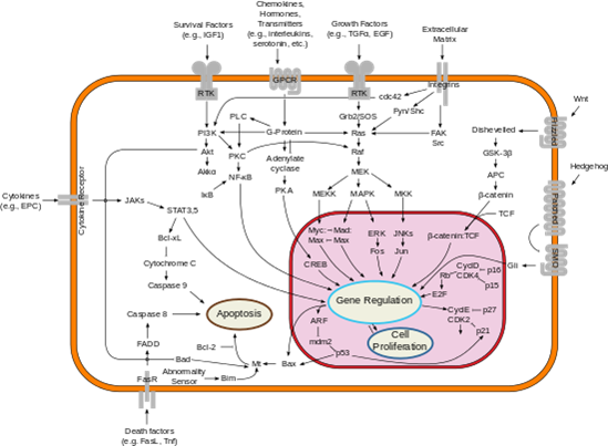
Bcl-2 gene (B-cell lymphoma-2) is a member of Bcl-2 family, which is the earliest discovered apoptosis protein. It is an anti-apoptosis protein containing multiple BH domains. It is highly expressed in some cancer cells and selectively plays an anti-tumor role. Bcl-2 family proteins play an important role in the regulation of apoptosis by forming dimers with Bax and dimerizing themselves. When Bcl-2 protein is inhibited, its dimer with Bax decreases, leading to cell apoptosis. When Bcl-2 protein is overexpressed, heterodimers formed by Bcl-2 protein and Bax increase and apoptosis is inhibited. The balance between Bcl-2 and Bax proteins at the cell death signal checkpoint determines the survival or apoptosis of cells.
Fig: Bcl-2 and Cell apoptosis
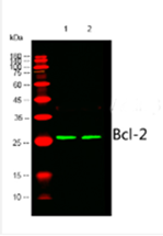
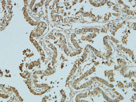
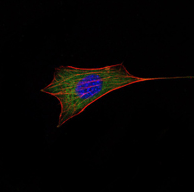
We recommend Abbkine selected Bcl-2 antibody for WB, IHC-P and IF experiments in human, rats, mice and chickens.
Common methods of cell proliferation mainly include MTT method, WST-1 method and CCK-8 method, which are mainly based on the activity of cells, thus reflecting the overall proliferation effect, but unable to detect individual proliferating cells. Recognized as the most accurate method to detect cell proliferation is to directly detect DNA synthesis in cells. Previously, BrdU method was the most commonly used method. However, the method requires DNA denaturation (such as acid denaturation, thermal denaturation or DNase digestion) to expose BrdU, thus binding to BrdU antibody. The whole experiment has many influencing factors and poor stability.
Abbkine innovates and develops EdU method based on EdU incorporation and subsequent click reaction to solve this application difficulty:
Product name
Cat#
Fluorescent characteristic
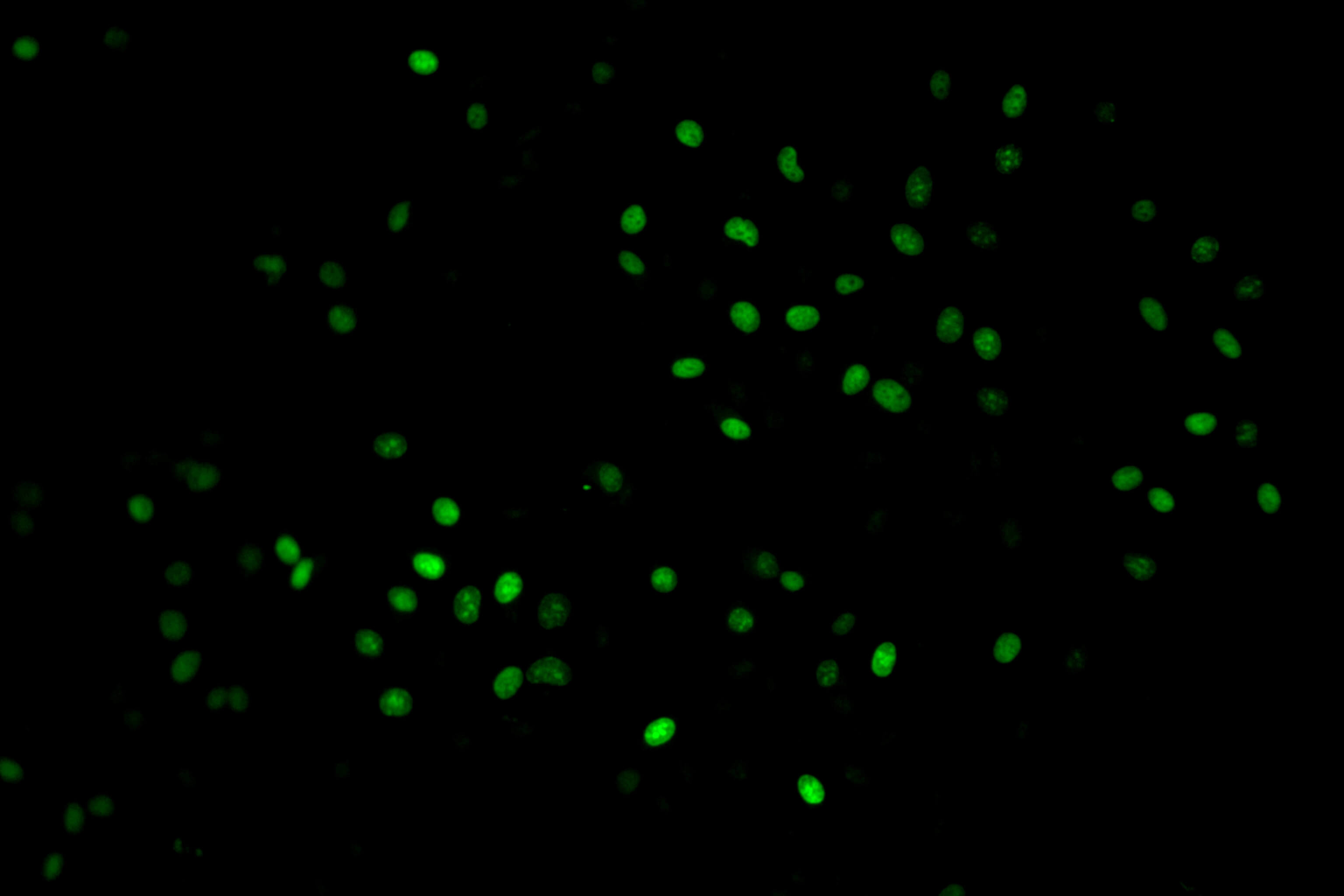
Cell Proliferation EdU Image Kit (Green Fluorescence)
Principle:EdU (5-ethynyl-2´-deoxyuridine) is a nucleoside analog of thymidine and is incorporated into DNA during active DNA synthesis. Comparing to BrdU assays, the EdU-Click Assays are not antibody based and therefore do not require DNA denaturation (typically using HCl or heat or digestion with DNase) for detection of the incorporated nucleoside. Detection is based on a click reaction, a copper-catalyzed covalent reaction between an azide and an alkyne, is complete within 30 minutes.
Kit components
Advantage
• EdU (10mM) • AbFluor 488 azide or AbFluor 545 azide • 10×Reaction buffer • Copper • Reducing Agent
• Not antibody based • No DNA denaturation. Can maintain cell morphology and DNA integrity • Patented AbFluor 488 and 545 azide have good light stability and quenching resistance • Optimization for fluorescence microscope
Promotion product list: Classical loading control and tag antibodies and the conjugated versions of these antibodies, including HRP, Biotin, FITC, Cy3, Cy5 and AbFlour TM dyes.
Details of activities: Buy 2 get 1 for free (if a single order is full of 3 antibodies, the antibody with the lowest price will be exempted).
Why choose Abbkine loading control and tag antibodies?
Abbkine offers a wide range of loading control and tag antibodies, including the hot targets and unique species, covering the molecular weights from 15 kDa to 116 kDa.
Abbkine conjugated loading control and tag antibodies have diverse conjugates, including enzyme and featured fluorescent dyes, with the application of WB, IHC, IF and IP experiments.
Abbkine loading control and tag antibodies are verified through rigorous procedures, popular with customers for their good performance and high sensitivity.
Different sizes for clients to choose, small size and bulk size are available, both with high cost-effectiveness.
Part of promotion product list:
Classical loading control and tag antibodies (Citation of over 100 SCI Documents)
IP experiment partner-Agarose/Magnetic beads conjugated tag antibodies
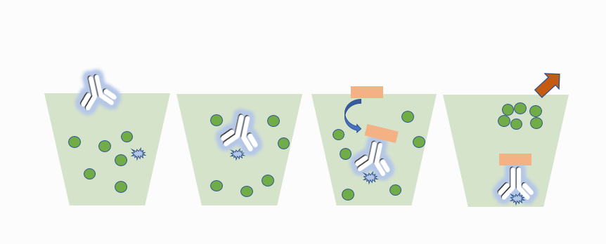
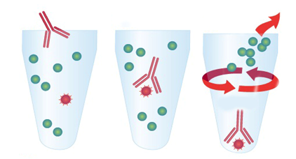
Compared with the traditional immunoprecipitation (IP) method using Protein A/G, Abbkine's agarose/magnetic bead coupling labeled antibody eliminates the step of Protein A/G binding with antigen-antibody complex. After the conjugated antibody is directly combined with the target protein, the target protein is separated from the cell lysate by centrifugation or using a magnetic rack. It not only makes the experiment simple and fast, but also can avoid non-specific binding and reduce the background.
For the full product promotion list, please contact Abbkine's local distributor or contact service@abbkine.com directly. Abbkine has the final right to interpret this activity.
Apoptosis is an orderly and programmed death followed by cells under the influence of physiological or pathological factors in order to maintain internal environment stability. Apoptosis occurs in cells. First, the cell volume shrinks and the connection disappears. Then, the density of cytoplasm increases, mitochondrial membrane potential disappears, permeability changes, cytochrome C is released to cytoplasm, nuclear substance is concentrated, nuclear membrane nucleolus is broken, DNA is degraded into fragments, and finally apoptotic bodies are formed and engulfed by macrophages.
Late apoptosis
In cell apoptosis, especially in the late stage of apoptosis, chromosomal DNA will be broken, resulting in a large number of sticky 3'-OH ends. Under the action of deoxynucleotide terminal transferase (TdT), derivatives formed by deoxyuridine triphosphate nucleotide (dUTP) and fluorescein can be labeled to the 3'- end of DNA, namely deoxynucleotide terminal transferase mediated nick end labeling (TUNEL). Normal or proliferating cells have few DNA breaks and can rarely be stained. Therefore, TUNEL is the most commonly used method to detect DNA fragmentation in late apoptosis.
Abbkine's TUNEL apoptosis detection kit provides a complete set of reagent components and optimized experimental scheme required for the experiment. It is suitable for a variety of instruments such as fluorescence enzyme labeling instrument, fluorescence microscope, flow cytometer, etc. It can be used to detect adherent cells, suspended cells, paraffin-embedded tissue sections, frozen sections and other sample types.
Apoptosis cannot be stopped once it starts, it is a highly regulated process.
Apoptosis can be initiated by two initial pathways. The internal initial pathway is activated by non-receptor stimulation, such as DNA damage, endoplasmic reticulum stress, metabolic stress, mitochondrial outer membrane permeability changes, such as cytochrome C release. The external pathway starts by binding death receptor to ligand (such as FasL, tumor necrosis factor TNF). After that, the two approaches converge in caspase cascade reaction, and cell death is induced by activating caspase protease or protein degrading enzyme. Anti-apoptotic ligands, such as cytokines and growth factors, promote cell survival, proliferation and differentiation through different signaling molecules such as AKT and p90RSK and anti-apoptotic proteins such as Bcl-2 and Bcl-x. The withdrawal of these cytokines and growth factors leads to cell death.
Abbkine has selected the antibody of apoptosis series. After multiple strict verifications, it is suitable for a variety of cell applications and meets the research needs of most customers. The excellent results are as follows:
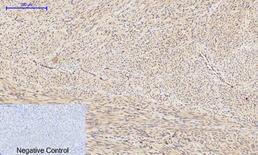
IHC analysis of paraffin-embedded human uterine tissue was performed using Bad polyclonal antibody diluted at 1: 200.
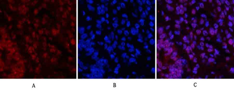
IF analysis of rat lung tissue was performed using Caspase-3 polyclonal antibody diluted at 1: 200.
Apoptosis is a dynamic process, which involves a series of complex biochemical reactions, expression regulation of multiple genes, signal transduction, cascade reactions involving multiple enzymes and multiple signal pathways. The cell feels the corresponding apoptosis signal stimulation, and a series of control switches in the cell are turned on or off, and activation of various enzymes triggers a series of cascade reactions. Different external factors initiate apoptosis in different ways, resulting in different signal transduction.
Early apoptosis
Feature 1: eversion of lipid membrane inside
In normal cells, phosphatidylserine (PS) is only distributed on the inner side of the lipid bilayer of the cell membrane. In the early stage of cell apoptosis, PS turns from the inner side of the lipid membrane to the outer side. Annexin V is a calcium-dependent phospholipid binding protein with a molecular weight of 35-36kD, and has high affinity with PS. It binds to the cell membrane of early apoptotic cells through PS exposed on the outside of cells. Therefore Annexin V is a sensitive indicator to detect early apoptosis of cells. Usually, Annexin V is labeled with some fluorescent probes, which can simply and directly detect PS eversion, an important feature of apoptosis.
AbFlourTM series of characteristic fluorescent dyes (blue, green and red) patented by Abbkine label recombinant human Annexin V protein, and are matched with PI to introduce different fluorescence apoptosis detection kits, which are suitable for detection with fluorescence equipment such as flow cytometry and fluorescence microscope.
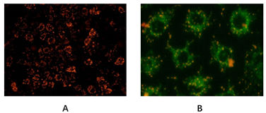
Normal mitochondrial membrane potential is the premise for maintaining mitochondrial oxidative phosphorylation and ATP production, and is necessary for maintaining mitochondrial function. The decline of mitochondrial membrane potential is a landmark event in the early stage of cell apoptosis. JC-1 is an ideal fluorescent probe widely used for detecting mitochondrial membrane potential. When mitochondrial membrane potential is high, JC-1 aggregates in the matrix of mitochondria to form polymers (J-aggregates). Red fluorescence can be generated(Ex/Em = 585/590). when the mitochondrial membrane potential is low, JC-1 cannot aggregate in the matrix of mitochondria. at this time, JC-1 is a monomer, which can generate green fluorescence (Ex/Em = 510/527 nm).
Abbkine Mitochondrial Membrane Potential Analysis Kit (JC-1) uses JC-1 as a fluorescent probe to rapidly and sensitively detect changes in mitochondrial membrane potential. This kit can very conveniently detect changes in mitochondrial membrane potential through changes in fluorescence color.
Step 1: Understand what immunoprecipitation is? Why should I do immunoprecipitation?
Immunoprecipitation (IP) is a method for purifying and enriching target proteins from specific mixtures (usually cell lysates or expression supernatants) by using antigen-antibody specific reactions. The traditional IP experiment is that after the antibody is combined with the target protein, it is incubated with agarose or magnetic beads coupled with Protein A /G (Fc fragment of binding antibody), and the bead-protein A/G- antibody-target protein complex is obtained by centrifugation. The complex is washed and eluted for subsequent downstream experiments. IP is an important step in many protein-related researches. It is used to study the existence, relative abundance, up-down regulation of protein expression, protein-to-stability and interaction of proteins.
Traditional IP protocol
Summary: The key tools I need to use in the whole process of IP are,
1) primary antibody: select the primary antibody that can react with the sample and do IP experiment;
Step 2: Finding that there is no suitable primary antibody to meet the immunoprecipitation I need to do?
Some target proteins cannot be immunoprecipitated because there is no corresponding specific antibody. For example, the antigen epitope recognized by the antibody is shielded by the interaction protein, unable to interact with the target protein or the antibody selection is inappropriate, the selected antibody cannot recognize the protein in natural conformation, etc. After an epitope tag protein is used, the tag antibody against this epitope can be selected for immunoprecipitation. Manufacturers will introduce some common label antibodies for IP experiments, involving labels such as flag, HA, His and Myc to meet the needs of most customers.
In particular, the agarose/magnetic bead coupled labeled antibody can optimize the immunoprecipitation experiment to the greatest extent and save time and cost.
1) Principle: The step of Protein A/G binding with antigen-antibody complex is omitted, and the problem of weak binding ability between Protein A/G and antibody is solved. After the conjugated antibody is directly combined with the target protein, the target protein is separated from the cell lysate by centrifugation or using a magnetic rack. The high-specificity monoclonal label antibody can realize high yield and high purity, and the stable and pre-sealed filler and the specific antibody reduce non-specific binding in the immunoprecipitation process.
Agarose/magnetic bead conjugated label antibody is applied to IP
Anti-Myc Tag Mouse Monoclonal Antibody (2D5), Magnetic Beads
Step 3: How to deal with heavy chain and light chain interference in WB (IP-WB) after immunoprecipitation?
In WB verification after immunoprecipitation experiment, when conventional Anti-IgG (H+L) enzyme labeled secondary antibody is used, two bands corresponding to heavy chain (50kDa) and light chain (25kDa) generated after denaturation of immunoprecipitation primary antibody will usually appear. If the molecular weight of the detected target protein is near here, it will be interfered by the two bands. How to avoid interference,
Method 1: Select the secondary antibody that only reacts with heavy chain or light chain. If the molecular weight of the target protein is less than 30KD, in order to avoid interference of antibody light chain, heavy chain specific secondary antibody is selected; If the molecular weight of the target protein is more than 30KD, in order to avoid interference of antibody heavy chain, light chain specific secondary antibody is selected;
Method 2: When selecting WB primary antibody, select different species from IP primary antibody to avoid interference from IP primary antibody;
Method 3: Select the WB primary antibody directly coupled with HRP, and omit the step of secondary antibody. Immune precipitation is broken in three steps.