2019年12月11日星期三

Selected primary antibody| Cleaved Caspase-3 antibody recommendation

Review of Related Articles:


Research
Hotspots of Cell Apoptosis: Bcl-2 Monoclonal Antibody


Abbkine
P53 antibody helps basic cancer research!


Marker
of myofibroblast antibody-α-SMA/ACTA2 Monoclonal Antibody


Selected
primary antibody|TNF-α antibody recommendation


Cleaved Caspase-3 is an Activated Form of
Caspase-3. Caspases are a family of cysteine proteases that are key mediators
of programmed cell death or apoptosis. The precursor form of all caspases is
composed of a prodomain, and large and small catalytic subunits. The active
forms of caspases are generated by several stimuli including ligand-receptor
interactions, growth factor deprivation and inhibitors of cellular functions.


All known caspases require cleavage
adjacent to aspartates to liberate one large and one small subunit, which
associate into a2b2 tetramer to form the active enzyme. Gene for Caspase-3 also
known as Yama, CPP32, and apopain codes for a 32-kDa protein. The main
substrate of Caspase-3 is poly (ADP- ribose) polymerase PARP (ADP-ribose) which
is related to DNA repair and gene integrity monitoring. When apoptosis is
initiated, Caspase-3 is activated by cleavage of Asp-28/Ser-29 (between
N-terminal prodomains) and Asp-175/Ser-176 (between large subunit and small
subunit), resulting in a large subunit of 17kDa and a small subunit of 12kDa.
P17 and p12 dimerize to form cleaved caspase-3 in activated form.
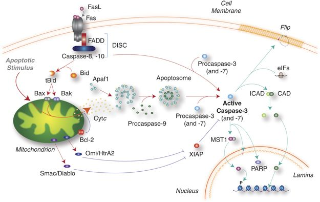


The picture is from https://www.nature.com/articles/cdd2009180.


AbbkineCleaved-Caspase-3
p17 (D175) Polyclonal Antibody helps basic cancer research! Basic information
about the product:


Product name

#CAT

Reactivity

Applications

Dilution ratio

Observed Band(KD)

Cleaved-Caspase-3 p17 (D175) Polyclonal
Antibody

ABP0021
human/mouse/rat
WB/IF/IHC-P/ELISA

WB (1:500-1:2000)
IF (1:50-1:300)
IHC-P (1:50-1:300)
ELISA(1:20000)

19/17 kD

Abbkine Cleaved-Caspase-3 p17 (D175)
Polyclonal Antibody verification results:
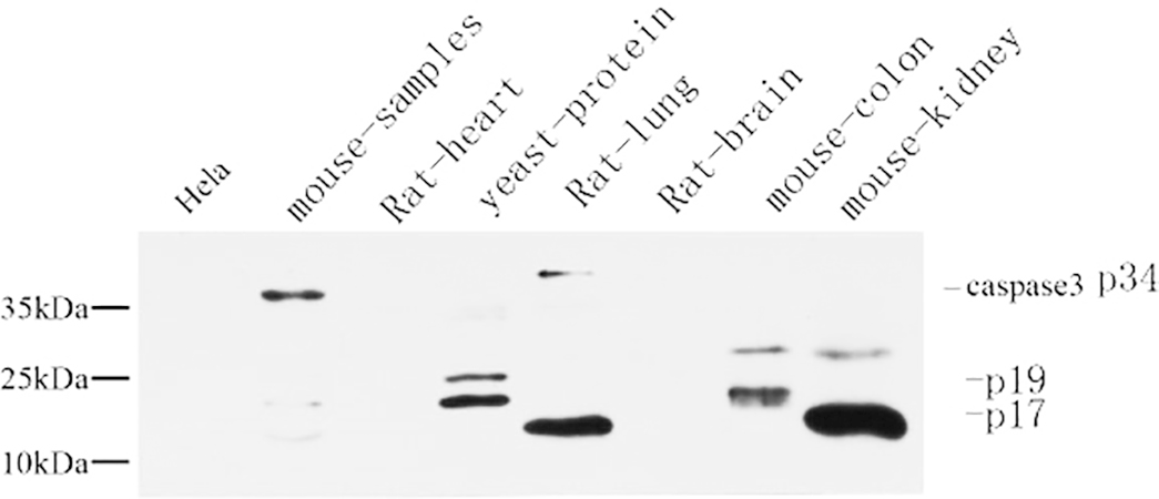


Western Blot analysis using Abbkine Cleaved-Caspase-3 p17 (D175) Polyclonal Antibody.


Immunohistochemical analysis of
paraffin-embedded mouse colon tissue (1:200) using Abbkine Cleaved-Caspase-3
p17 (D175) Polyclonal Antibody. (1:200).

Immunohistochemical analysis of
paraffin-embedded mouse colon tissue (1:200) using Abbkine Cleaved-Caspase-3
p17 (D175) Polyclonal Antibody. (1:200).

2019年11月27日星期三

Selected primary antibody|TNF-α antibody recommendation

Review of Related Articles:


Research
Hotspots of Cell Apoptosis: Bcl-2 Monoclonal Antibody


Abbkine
P53 antibody helps basic cancer research!


Marker of myofibroblast antibody-α-SMA/ACTA2 Monoclonal Antibody


Tumor necrosis factor (TNF, cachexin, or
cachectin; once named as tumor necrosis factor alpha or TNFα) is a cell
signaling protein (cytokine) involved in systemic inflammation and is one of
the cytokines that make up the acute phase reaction. It is produced chiefly by
activated macrophages, although it can be produced by many other cell types
such as CD4+ lymphocytes, NK cells, neutrophils, mast cells, eosinophils, and
neurons. TNF is a member of the TNF superfamily, consisting of various
transmembrane proteins with a homologous TNF domain.


The primary role of TNF is in the regulation of immune cells. TNF, being an endogenous pyrogen, is able to induce fever, apoptotic cell death, cachexia, inflammation and to inhibit tumorigenesis and viral replication and respond to sepsis via IL1- & IL6-producing cells. Dysregulation of TNF production has been implicated in a variety of human diseases including Alzheimer's disease, cancer, major depression, psoriasis and inflammatory bowel disease (IBD). Though controversial, studies of depression and IBD are currently being linked to increased levels of TNF. Recombinant TNF is used as an immunostimulant under the INN tasonermin. TNF can be produced ectopically in the setting of malignancy and parallels parathyroid hormone both in causing secondary hypercalcemia and in the cancers with which excessive production is associated.


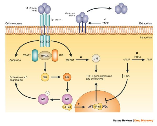
Fig: Anti-TNF-α therapies: the next generation (https://www.nature.com/articles/nrd1175)

Abbkine TNF-α
Polyclonal Antibody
helps basic cancer research! Basic information about
the product:


Product name

#CAT

Reactivity

Applications

Dilution ratio

Observed Band(KD)

TNF-α Polyclonal Antibody

ABP0127

Human/Mouse/Rat

WB/IF/IHC-P/ELISA

WB (1:500-1:2000)
IF (1:200-1:1000)
IHC-P (1:100-1:300)
ELISA(1/10000)

16KD

Abbkine TNF-α Polyclonal Antibody verification results:


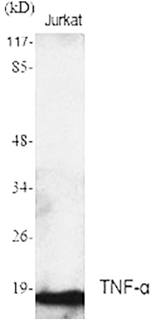
Western Blot analysis using Abbkine TNF-α Polyclonal Antibody.

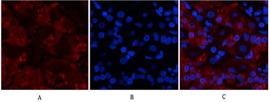
Immunofluorescence analysis of human
stomach tissue using Abbkine TNF-α Polyclonal Antibody (1:200).

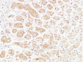
Immunohistochemical analysis of
paraffin-embedded Human stomach (1:100).

2019年11月14日星期四

Marker of myofibroblast antibody-α-SMA/ACTA2 Monoclonal Antibody

ACTA2 (actin alpha 2) is an actin protein
with several aliases including alpha-actin, alpha-actin-2, aortic smooth muscle
or alpha smooth muscle actin (α-SMA, SMactin, alpha-SM-actin, ASMA). Actins are
a family of globular multi-functional proteins that form microfilaments. ACTA2
is one of 6 different actin isoforms and is involved in the contractile
apparatus of smooth muscle. ACTA2 (as with all the actins) is extremely highly
conserved and found in nearly all mammals.


In humans, ACTA2 is encoded by the ACTA2
gene located on 10q22-q24. Mutations in this gene cause a variety of vascular
diseases, such as thoracic aortic disease, coronary artery disease, stroke,
Moyamoya disease, and multisystemic smooth muscle dysfunction syndrome.


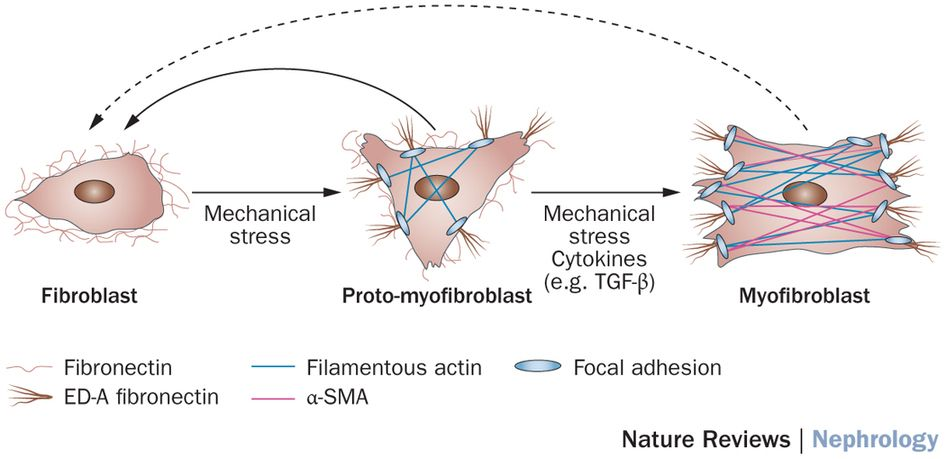
ACTA2 (commonly referred to as alpha-smooth muscle actin or α-SMA) is often used as a marker of myofibroblast formation.


Figure: Diverse origins of the
myofibroblast—implications for kidney fibrosis (From Nature Reviews)


Abbkine α-SMA Monoclonal Antibody Helps Basic Cancer Research! Basic information about the product:


Product name

#CAT

Reactivity

Applications

Dilution ratio

Observed Band(KD)

α-SMA Monoclonal
Antibody


ABM0052


Human/Mouse/ Rat

WB/IF/IHC-P

WB (1:10000-1:100000)
IF (1:100-1:200)
IHC-P (1:1000-1:2000)

42KD


Abbkine α-SMA Monoclonal Antibody verification results:


α-SMA-IF.png

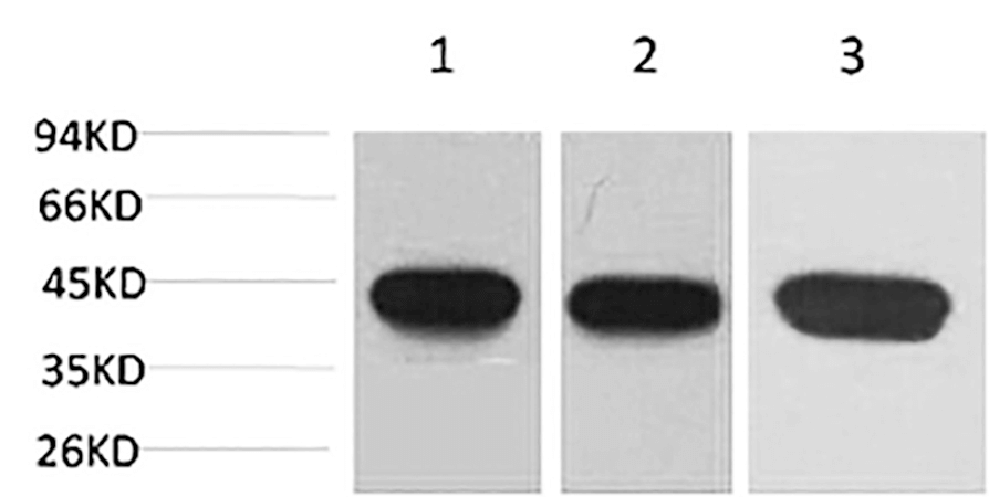
Western blot analysis of 1) Hela, 2) 3T3,
3) rat brain using α-SMA Monoclonal Antibody.
α-SMA-IHC

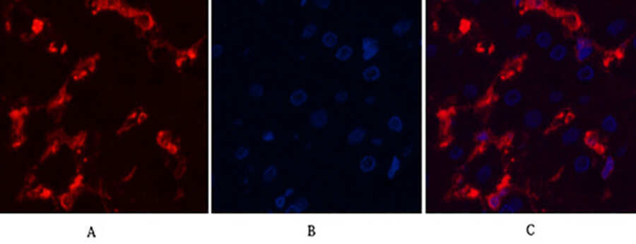
Immunofluorescence analysis of human
liver tissue.


α-SMA-WB

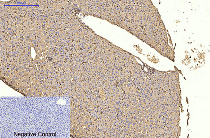
Immunohistochemical analysis of paraffin-embedded
mouse liver tissue

2019年11月4日星期一

Abbkine winter promotion: looking for you who need live-cell probe

Promotion time: From now to 15th January, 2020.


Promotion product
list:
TraKine™ Pro product
line (Tubulin, Lysosome, Mitochondrion, Nuclei live-cell probe ).


Details of activities: 20% off for TraKine™ Pro product line.


Details for promotion product:


Product name

#Cat

Target

Ex/Em (nm)

Size

Promotion price ($)

TraKine™ Pro Live-cell Tubulin Staining
kit (Green Fluorescence with Super Resolution)

KTC4100

Tubulin

500/520

50/250T

144/496

TraKine™ Pro Live-cell Lysosome Staining
kit (Deep Red Fluorescence with Super Resolution)

KTC4210

Lysosome

650/665

50/250T

128/472

TraKine™ Pro Live-cell Lysosome Staining
kit (Orange Fluorescence with Super Resolution)

KTC4220

Lysosome

565/590

50/250T

128/4721

TraKine™ Pro Live-cell Mitochondrion
Staining kit (Deep Red Fluorescence with Super Resolution)

KTC4300

Mitochondrion

647/661

50/250T

176/576

TraKine™ Pro Live-cell Nuclei Staining
kit (Deep Red Fluorescence with Super Resolution)

KTC4510

Nuclei

650/665

50/250T

144/496


TraKine™ Pro features & benefits:


  • Proprietary probe contains fluorescent dye and a unit which
    electively recognize organelles, with high specificity, low background,
    excellent photostability and good cell permeability.
  • Optimized staining protocol for labeling subcellular structures in
    mammalian living and fixed cells.
  • Fuorescence can last for several hours in cells, stable and
    persistent, especially ideal for monitoring dynamics.
  • Super resolution, especially suitable for Confocal and long-term
    super-resolution imaging (such as SIM, STED, TIRF, STORM and PALM).
  • Safe, very low or no cytotoxicity to cells.

2019年10月30日星期三

Abbkine P53 antibody helps basic cancer research!

P53 gene, human tumor suppressor gene. The gene encodes a protein with a molecular weight of 43.7KDa, but is named P53 because the protein band appears at 53KDa shown by Marker. Wild-type p53 causes cancer cells to apoptosis, thus preventing canceration; But also has the function of helping cell genes repair defects; Studies have found that mutant p53 can enhance canceration. 50% of human tumors are related to P53 gene. Currently, there are liver cancer, breast cancer, bladder cancer, gastric cancer, colon cancer, prostate cancer, soft tissue sarcoma, ovarian cancer, brain tumor, lymphocyte tumor, esophageal cancer, lung cancer, osteosarcoma, etc. P53 mutation in human tumors is mainly in highly conserved regions, with the highest mutation at 175, 248, 249, 273, 282 sites, and different types of tumors.  


P53 has a very short half-life, so it is almost undetectable in unstressed cells. This is mainly realized by the continuous ubiquitination of p53 and the degradation of 26S proteasome in the later stage under the action of its inhibitor MDM2. Cell stress signals such as DNA damage can inhibit ubiquitination of p53 and lead to its stabilization and activation. Once p53 is activated, it will bind to its downstream target regulatory elements and regulate its transcription. Therefore, various cell therapy schemes based on different tumor inhibition functions of p53 can be started. Just because past studies have shown that p53 gene mutations exist in about half of cancers, scientists regard them as important targets for developing new cancer therapies.


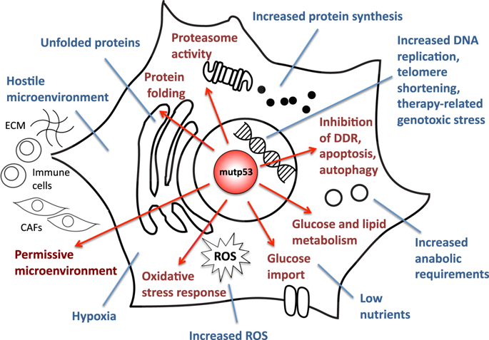
Mutant p53 promotes adaptive responses to cancer-related stress conditions to support tumor progression.

Abbkine P53 Antibody Helps Basic Cancer
Research! Basic information about the product:


Product name
#CAT
Reactivity Applications
Dilution ratio
p53 Polyclonal Antibody ABP0110   Human, Monkey, Mouse, Rat
WB/IF/IHC-P/ELISA

WB (1:500-1:2000)
IF (1:200-1:1000)
IHC-P (1:100-1:300)
ELISA  (1:10000).

Abbkine P53 Antibody verification results:


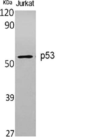
Western Blot analysis of various cells using p53 Polyclonal Antibody diluted at 1:1000.
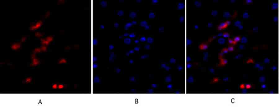
Immunofluorescence analysis of human liver tissue. 1, p53 Polyclonal Antibody (red) was diluted at 1:200 (4°C, overnight). 2, Cy3 Labeled secondary antibody was diluted at 1:300 (room temperature, 50min). 3, Picture B: DAPI (blue) 10min. Picture A: Target. Picture B: DAPI. Picture C: merge of A+B.
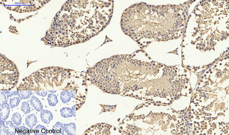
Immunohistochemical analysis of paraffin-embedded mouse testis tissue. 1, p53 Polyclonal Antibody was diluted at 1:200 (4°C, overnight). 2, Sodium citrate pH 6.0 was used for antibody retrieval (>98°C, 20min). 3, secondary antibody was diluted at 1:200 (room temperature, 30min). Negative control was used by secondary antibody only.

2019年10月15日星期二

IL-6: Target Molecule for Cytokine Storms

Interleukin-6 (IL-6) is a multi-effect cytokine
that plays an important role in host defense by regulating immune and
inflammatory reactions. IL-6 is mainly produced by macrophages, T lymphocytes
and B lymphocytes. When the body has inflammatory reaction or infection,
various cytokines such as virus, endotoxin and tumor necrosis factor can induce
the body to produce IL-6. The biological effects of IL-6 are mainly reflected
in inducing B cell differentiation to produce immunoglobulin, promoting T cell
proliferation and growth, enhancing blood cell differentiation and its
anti-tumor effect, promoting bone marrow hematopoietic stem cell proliferation,
etc.


IL-6 is an important member of cytokine network and plays a central role in acute inflammatory reaction. The production of IL-6 induces the production of C-reactive protein (CRP) and procalcitonin (PCT), which are directly related to inflammatory diseases and infection degree. IL-6 can diagnose early inflammation and warn the occurrence of sepsis faster. In addition, the half-life of IL-6 is shorter than CRP and PCT, which can reflect the effect of antibiotic treatment faster and better reflect the prognosis of patients. At the same time, IL-6 can be used as a biomarker of disease severity and prognosis indicators in cytokine storms, and its expression is superior to TNF-α and IL-1. A large number of research results show that IL-6 is a suitable target molecule for cytokine storms.


If you are studying IL-6 or are about to study IL-6, the recommended IL-6 cytokines are as follows:


Product
name


#cat

Sequence

Purity

Expressed
Host


Endotoxin

Formulation

Human
IL-6 protein

PRP100120

Human IL6
(NP_000591.1) (Val 30-Met 212)

>
95%

E.
coli

<1EU/μg

lyophilized
powder

Mouse IL-6 protein

PRP100371

Mouse
IL6 (P08505) (Phe25-Thr211)

>
95%

E.
coli

<1EU/μg

lyophilized
powder

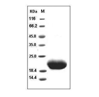
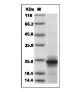
SDS-PAGE analysis:


Human IL-6 protein


Mouse IL-6 protein


Recommendations
for cytokine storage:


Cytokines
in lyophilized powder form are very stable and can be stored for several years
at -20℃ or -80℃. Freeze-dried powder should be dissolved before use and then
added to the culture medium in liquid form. The dissolved cytokines can only be
stored for a short time (about 1 week) at 4℃. For long-term preservation, first
prepare a diluent (which must contain carrier protein, such as 0.1% BSA, 5%HSA,
or 10% FBS), and then sub-pack and freeze at -20℃ or -80℃. Repeated freezing
and thawing must be avoided, as each freezing and thawing will cause partial
inactivation of proteins.

2019年10月10日星期四

Abbkine Meets You at 17th International Immunology Congress (IUIS 2019)

Abbkine
invites you to visit IUIS 2019, Beijing, China.


Abbkine featured products and services will be displayed at the exhibition. There always be one type can flash into your eyes. Participate in interaction, more surprises are waiting for you!


Date: October
19 – 23, 2019


Booth:#D22,level 4


Address: China
National Convention Center, Beijing, China


About: The 17th International Congress of Immunology (IUIS 2019) will be held from October 19 – 23, 2019 at the China National Convention Center, Beijing, China. IUIS 2019 promises to be an unforgettable event that will bring together delegates from all over the world. We anticipate over 5000 participants, including international leaders at the forefront of the discipline that will present the most recent advances in basic immunology and clinical translation.