Accurate protein quantification is necessary for protein-related experiments that involve research in molecular biology, cell biology, biochemistry, developmental biology, and neuroscience. Abbkine protein quantification products include Protein Quantification Kits (BCA Assay and Bradford Assay), SuperLumia ECL HRP Substrate Kit, Protein Gel Flash Staining Kit, Colorcode Prestained Protein Marker and so on.
Protein Quantification Kits
Featured products:
1. Protein Quantification Kits (BCA Assay)
• Save time—Easier and 4x faster than traditional Lowry methods.
• High sensitivity—The detection line is as low as 25ug/ml, and the minimum detection protein is 0.5ug.
• Good compatibility—unaffected by most ionic and non-ionic detergents. Compatible with up to 5% SDS, 5% Triton X-100, 5% Tween-20, 60, 80 in the sample.
• Excellent linearity-linear standard curve range: 50– 1000ug/ml.
• Good stability—Protein-to-protein differences are smaller compared to dye-bound methods.
2. Protein Quantification Kits (Bradford Assay)
• Save time-rapid color development.
• Good compatibility—unaffected by most chemicals. The compatible concentrations of mercaptoethanol and dithiothreitol in the samples can reach 1mM and 5mM, respectively.
• Good detection range—The detection range is 50-1000ug/ml.
ECL
SuperLumia ECL HRP Substrate Kit developed by Abbkine is based on the classic peroxidase substrate system innovation and optimization. It is used to enhance chemiluminescence (ECL) and can directly replace other expensive ECL products without re-optimizing experimental conditions. It is the perfect combination of superior quality and affinity price.
Protein Gel Flash Staining Kit
Protein staining can be detected in the range of 10ng to 5μg, which only takes 0.5-1hours. It is about 5-10 times more sensitive than the traditional Coomassie R-250 dye.
Fig1. Various volumes of BSA samples were separated on 10% SDS-PAGE, stained with protein gel flash staining working solution for 30 minutes.
Protein Marker
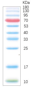
Abbkine's Colorcode Prestained Protein Marker (10-180 kDa) contains 3 colors, totaling 10 protein standards (10, 17, 25, 33, 40, 53, 70, 95, 130, 180 kDa). The orange band is 70 kDa; the green band is 10 kDa. Thaw at room temperature. Mix gently and thoroughly to ensure a homogeneous solution.
Fig2. Image is from a 15% Tris-glycine gel (SDS-PAGE) transferred to membrane using Abbkine Colorcode Prestained Protein Marker (10-180 kDa).
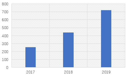
Since Abbkine document collection began, as of December 31, 2019, the number of English articles published by google using Abbkine products has exceeded 1400, with an impact factor exceeding 5400 points.
Thank you for your trust and support to Abbkine. We will continuously stimulate our internal creativity, provide competitive biomedical products and services, and continuously create maximum value for our customers. With a view to becoming a respected and world-class provider of biomedical products and services.
Figure 1: Number of English Articles Published Using Abbkine Products from 2017 to 2019
In December 2019, Abbkine added 200+ citations. Some high-score citations are as below.
1.LECT2, a Ligand for Tie1, Plays a Crucial Role in Liver Fibrogenesis.
Abstract: Liver fibrosis is a very common condition seen in millions of patients with various liver diseases, and yet no effective treatments are available owing to poorly characterized molecular pathogenesis. Here, we show that leukocyte cell-derived chemotaxin 2 (LECT2) is a functional ligand of Tie1, a poorly characterized endothelial cell (EC)-specific orphan receptor. Upon binding to Tie1, LECT2 interrupts Tie1/Tie2 heterodimerization, facilitates Tie2/Tie2 homodimerization, activates PPAR signaling, and inhibits the migration and tube formations of EC. In vivo studies showed that LECT2 overexpression inhibits portal angiogenesis, promotes sinusoid capillarization, and worsens fibrosis, whereas these changes were reversed in Lect2-KO mice. Adeno-associated viral vector serotype 9 (AAV9)-LECT2 small hairpin RNA (shRNA) treatment significantly attenuates fibrosis. Upregulation of LECT2 is associated with advanced human liver fibrosis staging. We concluded that targeting LECT2/Tie1 signaling may represent a potential therapeutic target for liver fibrosis, and serum LECT2 level may be a potential biomarker for the screening and diagnosis of liver fibrosis.
Abstract: In flowering plants, EMS1 (Excess Microsporocytes 1) perceives TPD1 (Tapetum Determinant 1) to specify tapeta, the last somatic cell layer nurturing pollen development. However, the signaling components downstream of EMS1 are relatively unknown. Here, we use a molecular complementation approach to investigate the downstream components in EMS1 signaling. We show that the EMS1 intracellular domain is functionally interchangeable with that of the brassinosteroid receptor BRI1 (Brassinosteroid Insensitive 1). Furthermore, expressing EMS1 together with TPD1 in the BRI1 expression domain could partially rescue bri1 phenotypes, and led to the dephosphorylation of BES1, a hallmark of active BRI1 signaling. Conversely, expressing BRI1 in the EMS1 expression domain could partially rescue ems1 phenotypes. We further show that PpEMS1 and PpTPD1 from the early land plant Physcomitrella patens could completely rescue ems1 and tpd1 phenotypes, respectively. We propose that EMS1 and BRI1 have evolved distinct extracellular domains to control different biological processes but can act via a common intracellular signaling pathway.
Abstract: The innate immune sensor NLRP3 assembles an inflammasome complex with NEK7 and ASC to activate caspase‐1 and drive the maturation of proinflammatory cytokines IL‐1β and IL‐18. NLRP3 inflammasome activity must be tightly controlled, as its over‐activation is involved in the pathogenesis of inflammatory diseases. Here, we show that NLRP3 inflammasome activation is suppressed by a centrosomal protein Spata2. Spata2 deficiency enhances NLRP3 inflammasome activity both in the macrophages and in an animal model of peritonitis. Mechanistically, Spata2 recruits the deubiquitinase CYLD to the centrosome for deubiquitination of polo‐like kinase 4 (PLK4), the master regulator of centrosome duplication. Deubiquitination of PLK4 facilitates its binding to and phosphorylation of NEK7 at Ser204. NEK7 phosphorylation in turn attenuates NEK7 and NLRP3 interaction, which is required for NLRP3 inflammasome activation. Pharmacological or shRNA‐mediated inhibition of PLK4, or mutation of the NEK7 Ser204 phosphorylation site, augments NEK7 interaction with NLRP3 and causes increased NLRP3 inflammasome activation. Our study unravels a novel centrosomal regulatory pathway of inflammasome activation and may provide new therapeutic targets for the treatment of NLRP3‐associated inflammatory diseases.
Products using from Abbkine:
IFKine™ Green Donkey Anti-Mouse IgG (CAT#: A24211)
4. Cross-Microbial Protection via Priming a Conserved Immune Co-Receptor through Juxtamembrane Phosphorylation in Plants
Abstract: Living organisms can be primed for potentiated responses to recurring stresses based on prior experience. However, the molecular basis of immune priming remains elusive in plants that lack adaptive immunity. Here, we report that bacterial challenges can prepare plants for fungal attacks by inducing juxtamembrane phosphorylation of CERK1, the co-receptor indispensable for signaling in response to the fungal elicitor chitin. This phosphorylation is mediated by BAK1, a co-receptor for signaling in response to multiple elicitors. BAK1 interacts with CERK1, and loss of BAK1 reduces priming phosphorylation of CERK1. Juxtamembrane phosphomimetic mutations of CERK1 confer accelerated chitin responses and fortified fungal resistance without triggering constitutive immunity, whereas juxtamembrane phosphodeficient mutations diminish bacteria-induced protection against fungal infection. These findings reveal that crosstalk between cell-surface immune co-receptors can prime defense and demonstrate that juxtamembrane phosphorylation of plant receptor-like kinases can occur independent of kinase activation to place the protein into a prime state.
Products using from Abbkine:
Anti-GST Tag Mouse Monoclonal Antibody (2A8) (CAT#: A02030)
5. Extracellular vesicles of carcinoma-associated fibroblasts creates a pre-metastatic niche in the lung through activating fibroblasts
Abstract: Carcinoma-associated fibroblasts (CAFs) have been known to promote cancer progression by modifying the primary tumor microenvironment. We aimed to elucidate the intercellular communication between CAFs and secondary organs in salivary adenoid cystic carcinoma (SACC) metastasis.
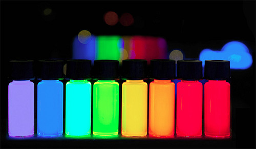
AbFluor™ dyes are a series of highly water-soluble fluorescent dyes introduced by Abbkine after many years of research, covering the ultraviolet, visible and near-infrared spectrum, suitable for labeling biological macromolecules, especially proteins and nucleic acids. Their excellent hydrophilicity enables protein-conjugated labeling to be performed easily in aqueous media, while minimizing the need for organic solvents. AbFluor™ dyes not only have better fluorescence performance than other fluorescent dyes (such as FITC, TRITC, Alexa Fluor and Dylight dyes), but also the coupling products are more stable.
Why recommend AbFluor™ fluorescent dyes?
With innovative modifications to the core structure of the dye, AbFluor™ dyes have more brilliant properties than other commercial dyes, including stronger fluorescent brightness, better light stability, wider pH tolerance range, and better penetration and lower background. As a third-generation fluorescent dye, AbFluor™ dyes not only perform better than traditional dyes (such as FITC, TRITC, and Cy dyes), but also perform better than other second-generation commercial dyes (such as Alexa Fluor, Dylight and IRDye).
1. AbFluor™ dyes have excellent core structure, stable reactive groups, and good light stability, ensuring high stability and high labeling efficiency of bioconjugation.
2. High water solubility (>100 mg/ml), which minimizes fluorescence quenching; it is also easily soluble in other polar solvents, such as DMSO, DMF, methanol and ethanol.
3. A wide range of fluorescence spectrum, covering ultraviolet, visible, and near-infrared, to meet most of the experimental needs of researchers.
4. Good pH tolerance, stable in pH 2-11.
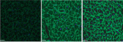
Taking AbFluor™ 488 as an example, the following is an IF comparison chart of different dye effects:
Different dye : FITC Alexa Fluor 488 AbFluor™ 488
So, are you excited, and what are you waiting for???
Western Blot is an experimental method commonly used in molecular biology, biochemistry and immunogenetics. The basic principle is to color the cell or biological tissue samples treated by gel electrophoresis through specific antibodies. Information on the expression of specific proteins in the analyzed cells or tissues is obtained by analyzing the staining position and staining depth.
Sample preparation:
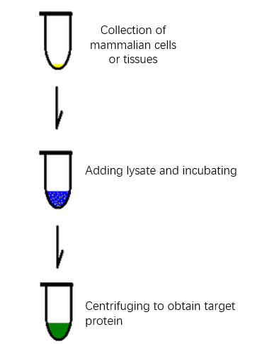
Good protein is half the success of WB. We need to lyse cells and tissues to extract the target protein. Once cracking occurs, hydrolysis, dephosphorylation and denaturation begin. If appropriate protease or phosphatase inhibitor is added to the cracking solution, these reactions will be greatly slowed down and the chance of extracting good protein will be increased.
It takes time and effort to prepare lysis solution by oneself, and the whole experiment may be wasted due to improper preparation. Why not try the protein extraction kit that Abbkine has packed for you? The kit is equipped with optimized lysis components. You only need to carry out the experiment according to the experimental steps.
High purity and no pollution-the extracted protein has high purity and maintains natural activity, reducing cross contamination;
Strong timeliness-non-denatured active protein can be purified within two hours;
Compatible application-the extracted protein can be directly used in subsequent proteomics applications;
Follow-up worry-free-kits own a special protease inhibitor package, anti-degradation, anti-useless;
High cost performance.
Characteristics and Advantages of Abbkine Protease Inhibitor Cocktail (100X):
Abbkine Protease Inhibitor Cocktail is an universal protease inhibitor cocktail contains individual components, including AEBSF, Aprotinin, Bestatin, E-64, Leupeptin and Pepstatin A with a broad specificity for cysteine, serine, acid proteases, and aminopeptidases. This protease inhibitor cocktail has been optimized and tested for mammalian cells and tissue extracts.
You deserve to have good sample preparation products!
Thank you all for giving our company full support in 2019. With the approaching of 2020 Spring Festival, here Abbkine makes the following holiday arrangement:
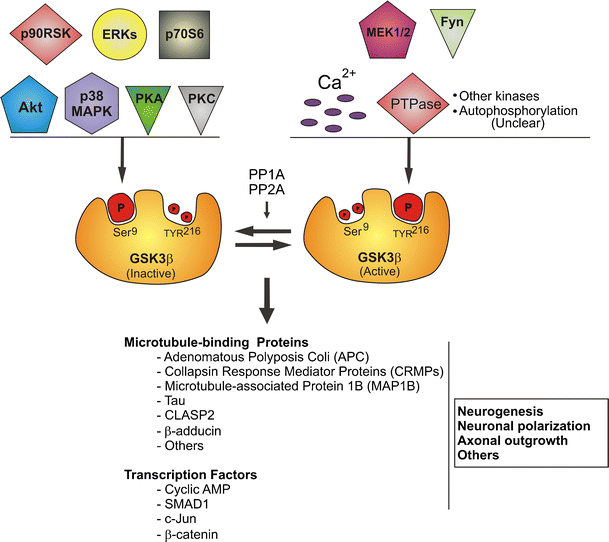
Glycogen synthase kinase-3 (GSK-3) is a serine-threonine kinase that has been evolutionarily very conservative.The protein encoded by GSK3β (glycogen synthase kinase 3 beta) is a serine-threonine kinase, belonging to the glycogen synthase kinase subfamily. It is involved in energy metabolism, neuronal cell development, and body pattern formation.
GSK-3β can also act on many signaling protein structural proteins and transcription factors, and regulate cell differentiation, proliferation, survival and apoptosis. GSK-3β is a key enzyme involved in liver glucose metabolism. It inhibits its activity by phosphorylating GS, reduces liver glucose synthesis, increases blood glucose concentration in the body, is controlled by insulin in the insulin signaling pathway, and participates in liver glucose metabolism.
Polymorphisms in GSK3β have been implicated in modifying risk of Parkinson disease, and studies in mice show that overexpression of GSK3β may be relevant to the pathogenesis of Alzheimer disease. Alternatively spliced transcript variants encoding different isoforms have been found for GSK3β .
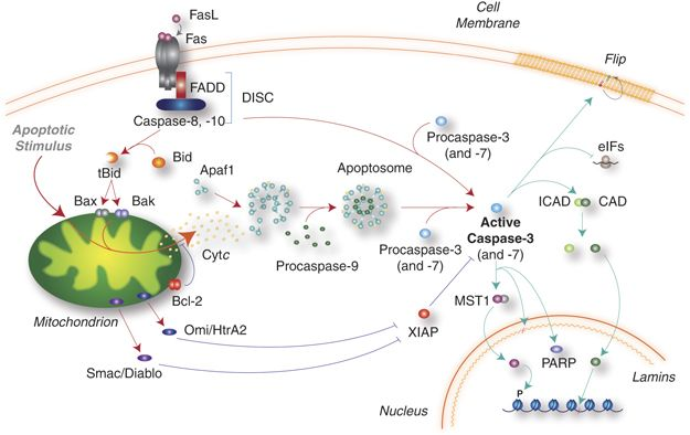
Cleaved Caspase-3 is an Activated Form of Caspase-3. Caspases are a family of cysteine proteases that are key mediators of programmed cell death or apoptosis. The precursor form of all caspases is composed of a prodomain, and large and small catalytic subunits. The active forms of caspases are generated by several stimuli including ligand-receptor interactions, growth factor deprivation and inhibitors of cellular functions.
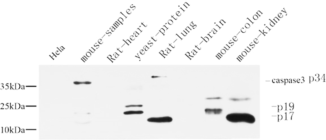
All known caspases require cleavage adjacent to aspartates to liberate one large and one small subunit, which associate into a2b2 tetramer to form the active enzyme. Gene for Caspase-3 also known as Yama, CPP32, and apopain codes for a 32-kDa protein. The main substrate of Caspase-3 is poly (ADP- ribose) polymerase PARP (ADP-ribose) which is related to DNA repair and gene integrity monitoring. When apoptosis is initiated, Caspase-3 is activated by cleavage of Asp-28/Ser-29 (between N-terminal prodomains) and Asp-175/Ser-176 (between large subunit and small subunit), resulting in a large subunit of 17kDa and a small subunit of 12kDa. P17 and p12 dimerize to form cleaved caspase-3 in activated form.