2019年12月24日星期二

Western Blot Product Portfolio- Sample Preparation

Overview of western blot (WB):


Western Blot is an experimental method
commonly used in molecular biology, biochemistry and immunogenetics. The basic
principle is to color the cell or biological tissue samples treated by gel
electrophoresis through specific antibodies. Information on the expression of
specific proteins in the analyzed cells or tissues is obtained by analyzing the
staining position and staining depth.


Sample preparation:


Good protein is half the success of WB. We
need to lyse cells and tissues to extract the target protein. Once cracking
occurs, hydrolysis, dephosphorylation and denaturation begin. If appropriate
protease or phosphatase inhibitor is added to the cracking solution, these
reactions will be greatly slowed down and the chance of extracting good protein
will be increased.


It takes time and effort to prepare lysis
solution by oneself, and the whole experiment may be wasted due to improper
preparation. Why not try the protein extraction kit that Abbkine has packed for
you? The kit is equipped with optimized lysis components. You only need to
carry out the experiment according to the experimental steps.


Abbkine sample
preparation product recommendation:


Product name

#Cat

Szie

Price ($)

ExKine™ Nuclear
and Cytoplasmic Protein Extraction Kit

KTP3001

50/200T

90/220

ExKine™ Nuclear
Protein Extraction Kit

KTP3002

50/200T

80/200

ExKine™
Cytoplasmic Protein Extraction Kit

KTP3003

50/200T

80/200

ExKine™ Total
Membrane Protein Extraction Kit

KTP3004

50/200T

80/200

ExKine™ Membrane
and Cytoplasmic Protein Extraction Kit

KTP3005

50/200T

90/220

ExKine™ Total
Protein Extraction Kit

KTP3006

50/200T

80/200

Protease
Inhibitor Cocktail (100X)

BMP1001

1ml/1mlx5

40/120


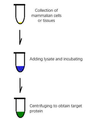
The basic process of protein extraction


Characteristics and Advantages of Abbkine
Protein Extraction Kit:


  • High purity and no pollution-the extracted protein has high purity and maintains natural activity, reducing cross contamination;
  • Strong timeliness-non-denatured active protein can be purified within two hours;
  • Compatible application-the extracted protein can be directly used in subsequent proteomics applications;
  • Follow-up worry-free-kits own a special protease inhibitor package, anti-degradation, anti-useless;
  • High cost performance.

Characteristics and Advantages of Abbkine Protease Inhibitor
Cocktail (100X):


Abbkine Protease Inhibitor Cocktail is an
universal protease inhibitor cocktail contains individual components, including
AEBSF, Aprotinin, Bestatin, E-64, Leupeptin and Pepstatin A with a broad
specificity for cysteine, serine, acid proteases, and aminopeptidases. This
protease inhibitor cocktail has been optimized and tested for mammalian cells
and tissue extracts.


You deserve to have good sample preparation
products!
2019年12月23日星期一

Abbkine Holiday Arrangement of 2020 Spring Festival

Dear partners:


Thank you all for giving our
company full support in 2019. With the approaching of 2020 Spring Festival,
here Abbkine makes the following holiday arrangement:


Holiday Time: 20th
Jan. 2020-31th Jan. 2020 (12 Days).


Holiday Time for the
logistics department: 18th Jan. 2020-2nd Feb. 2020 (16
Days).


Sorry for the inconvenience
caused by the holiday. My sincere regards.


Abbkine Scientific Inc


23th Dec.2019

2019年12月19日星期四

Selected primary antibody| GSK3β Mouse Monoclonal antibody recommendation

Review of Related Articles:


Research
Hotspots of Cell Apoptosis: Bcl-2 Monoclonal Antibody


Abbkine
P53 antibody helps basic cancer research!


Marker
of myofibroblast antibody-α-SMA/ACTA2 Monoclonal Antibody


Selected
primary antibody|TNF-α antibody recommendation


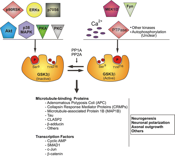
Glycogen synthase kinase-3 (GSK-3) is a
serine-threonine kinase that has been evolutionarily very conservative.The
protein encoded by GSK3β (glycogen synthase kinase 3 beta) is a
serine-threonine kinase, belonging to the glycogen synthase kinase subfamily.
It is involved in energy metabolism, neuronal cell development, and body
pattern formation.


GSK-3β can also act on many signaling
protein structural proteins and transcription factors, and regulate cell
differentiation, proliferation, survival and apoptosis. GSK-3β is a key enzyme
involved in liver glucose metabolism. It inhibits its activity by
phosphorylating GS, reduces liver glucose synthesis, increases blood glucose
concentration in the body, is controlled by insulin in the insulin signaling
pathway, and participates in liver glucose metabolism.


Polymorphisms in GSK3β have been implicated in modifying risk of Parkinson disease, and studies in mice show that overexpression of GSK3β may be relevant to the pathogenesis of Alzheimer disease. Alternatively spliced transcript variants encoding different isoforms have been found for GSK3β .


The picture is from https://link.springer.com/article/10.1007/s12035-013-8571-y


AbbkineGSK3β Mouse Monoclonal antibody (1A6) helps basic research! Basic information about the product:


Product name

#CAT

Reactivity

Applications

Dilution ratio

Observed Band(KD)

GSK3β Mouse Monoclonal antibody (1A6)

ABM0076
human/mouse/rat
WB/IHC-P
WB (1:1000-1:2000) IHC-P (1:100-1:200)
46 kD

GSK3β Mouse Monoclonal antibody (1A6)
verification results:


Immunohistochemical analysis of paraffin-embedded Human Breast Carcinoma Tissue using GSK3β Mouse mAb diluted at 1:200.

Immunohistochemical analysis of paraffin-embedded Human Stomach Carcinoma Tissue using GSK3β Mouse mAb diluted at 1:200.

Western blot analysis of 1) Hela Cell Lysate, 2) 3T3 Cell Lysate, 3) Rat Brain Tissue Lysate using GSK3β Mouse mAb diluted at 1:1000.

2019年12月11日星期三

Selected primary antibody| Cleaved Caspase-3 antibody recommendation

Review of Related Articles:


Research
Hotspots of Cell Apoptosis: Bcl-2 Monoclonal Antibody


Abbkine
P53 antibody helps basic cancer research!


Marker
of myofibroblast antibody-α-SMA/ACTA2 Monoclonal Antibody


Selected
primary antibody|TNF-α antibody recommendation


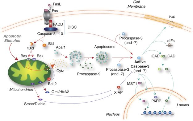
Cleaved Caspase-3 is an Activated Form of
Caspase-3. Caspases are a family of cysteine proteases that are key mediators
of programmed cell death or apoptosis. The precursor form of all caspases is
composed of a prodomain, and large and small catalytic subunits. The active
forms of caspases are generated by several stimuli including ligand-receptor
interactions, growth factor deprivation and inhibitors of cellular functions.


All known caspases require cleavage
adjacent to aspartates to liberate one large and one small subunit, which
associate into a2b2 tetramer to form the active enzyme. Gene for Caspase-3 also
known as Yama, CPP32, and apopain codes for a 32-kDa protein. The main
substrate of Caspase-3 is poly (ADP- ribose) polymerase PARP (ADP-ribose) which
is related to DNA repair and gene integrity monitoring. When apoptosis is
initiated, Caspase-3 is activated by cleavage of Asp-28/Ser-29 (between
N-terminal prodomains) and Asp-175/Ser-176 (between large subunit and small
subunit), resulting in a large subunit of 17kDa and a small subunit of 12kDa.
P17 and p12 dimerize to form cleaved caspase-3 in activated form.


The picture is from https://www.nature.com/articles/cdd2009180.


AbbkineCleaved-Caspase-3
p17 (D175) Polyclonal Antibody helps basic cancer research! Basic information
about the product:


Product name

#CAT

Reactivity

Applications

Dilution ratio

Observed Band(KD)

Cleaved-Caspase-3 p17 (D175) Polyclonal
Antibody

ABP0021
human/mouse/rat
WB/IF/IHC-P/ELISA

WB (1:500-1:2000)
IF (1:50-1:300)
IHC-P (1:50-1:300)
ELISA(1:20000)

19/17 kD

Abbkine Cleaved-Caspase-3 p17 (D175)
Polyclonal Antibody verification results:


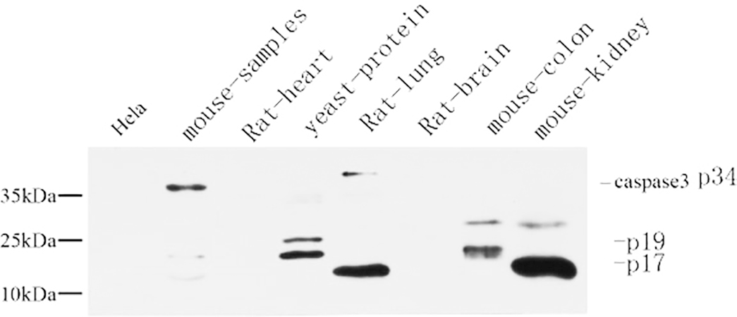
Western Blot analysis using Abbkine Cleaved-Caspase-3 p17 (D175) Polyclonal Antibody.


Immunohistochemical analysis of
paraffin-embedded mouse colon tissue (1:200) using Abbkine Cleaved-Caspase-3
p17 (D175) Polyclonal Antibody. (1:200).

Immunohistochemical analysis of
paraffin-embedded mouse colon tissue (1:200) using Abbkine Cleaved-Caspase-3
p17 (D175) Polyclonal Antibody. (1:200).

2019年11月27日星期三

Selected primary antibody|TNF-α antibody recommendation

Review of Related Articles:


Research
Hotspots of Cell Apoptosis: Bcl-2 Monoclonal Antibody


Abbkine
P53 antibody helps basic cancer research!


Marker of myofibroblast antibody-α-SMA/ACTA2 Monoclonal Antibody


Tumor necrosis factor (TNF, cachexin, or
cachectin; once named as tumor necrosis factor alpha or TNFα) is a cell
signaling protein (cytokine) involved in systemic inflammation and is one of
the cytokines that make up the acute phase reaction. It is produced chiefly by
activated macrophages, although it can be produced by many other cell types
such as CD4+ lymphocytes, NK cells, neutrophils, mast cells, eosinophils, and
neurons. TNF is a member of the TNF superfamily, consisting of various
transmembrane proteins with a homologous TNF domain.


The primary role of TNF is in the regulation of immune cells. TNF, being an endogenous pyrogen, is able to induce fever, apoptotic cell death, cachexia, inflammation and to inhibit tumorigenesis and viral replication and respond to sepsis via IL1- & IL6-producing cells. Dysregulation of TNF production has been implicated in a variety of human diseases including Alzheimer's disease, cancer, major depression, psoriasis and inflammatory bowel disease (IBD). Though controversial, studies of depression and IBD are currently being linked to increased levels of TNF. Recombinant TNF is used as an immunostimulant under the INN tasonermin. TNF can be produced ectopically in the setting of malignancy and parallels parathyroid hormone both in causing secondary hypercalcemia and in the cancers with which excessive production is associated.


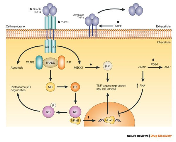
Fig: Anti-TNF-α therapies: the next generation (https://www.nature.com/articles/nrd1175)

Abbkine TNF-α
Polyclonal Antibody
helps basic cancer research! Basic information about
the product:


Product name

#CAT

Reactivity

Applications

Dilution ratio

Observed Band(KD)

TNF-α Polyclonal Antibody

ABP0127

Human/Mouse/Rat

WB/IF/IHC-P/ELISA

WB (1:500-1:2000)
IF (1:200-1:1000)
IHC-P (1:100-1:300)
ELISA(1/10000)

16KD

Abbkine TNF-α Polyclonal Antibody verification results:


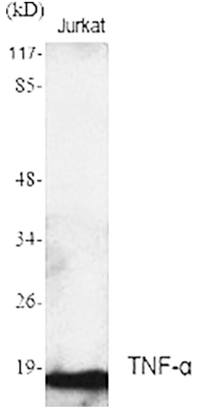
Western Blot analysis using Abbkine TNF-α Polyclonal Antibody.

Immunofluorescence analysis of human
stomach tissue using Abbkine TNF-α Polyclonal Antibody (1:200).

Immunohistochemical analysis of
paraffin-embedded Human stomach (1:100).

2019年11月14日星期四

Marker of myofibroblast antibody-α-SMA/ACTA2 Monoclonal Antibody

ACTA2 (actin alpha 2) is an actin protein
with several aliases including alpha-actin, alpha-actin-2, aortic smooth muscle
or alpha smooth muscle actin (α-SMA, SMactin, alpha-SM-actin, ASMA). Actins are
a family of globular multi-functional proteins that form microfilaments. ACTA2
is one of 6 different actin isoforms and is involved in the contractile
apparatus of smooth muscle. ACTA2 (as with all the actins) is extremely highly
conserved and found in nearly all mammals.


In humans, ACTA2 is encoded by the ACTA2
gene located on 10q22-q24. Mutations in this gene cause a variety of vascular
diseases, such as thoracic aortic disease, coronary artery disease, stroke,
Moyamoya disease, and multisystemic smooth muscle dysfunction syndrome.


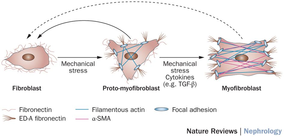
ACTA2 (commonly referred to as alpha-smooth muscle actin or α-SMA) is often used as a marker of myofibroblast formation.


Figure: Diverse origins of the
myofibroblast—implications for kidney fibrosis (From Nature Reviews)


Abbkine α-SMA Monoclonal Antibody Helps Basic Cancer Research! Basic information about the product:


Product name

#CAT

Reactivity

Applications

Dilution ratio

Observed Band(KD)

α-SMA Monoclonal
Antibody


ABM0052


Human/Mouse/ Rat

WB/IF/IHC-P

WB (1:10000-1:100000)
IF (1:100-1:200)
IHC-P (1:1000-1:2000)

42KD


Abbkine α-SMA Monoclonal Antibody verification results:


α-SMA-IF.png

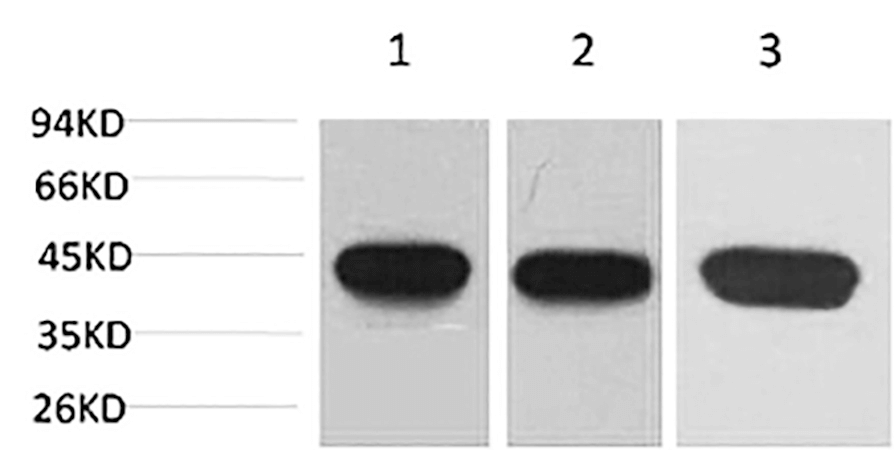
Western blot analysis of 1) Hela, 2) 3T3,
3) rat brain using α-SMA Monoclonal Antibody.
α-SMA-IHC

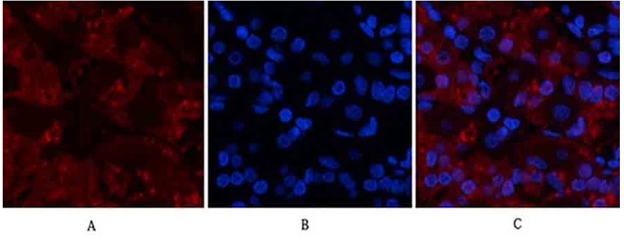
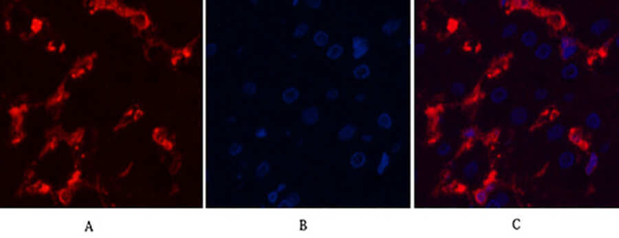
Immunofluorescence analysis of human
liver tissue.


α-SMA-WB

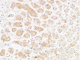
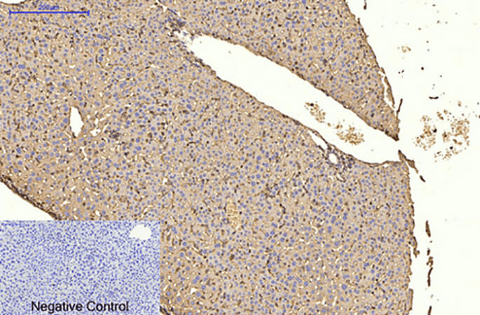
Immunohistochemical analysis of paraffin-embedded
mouse liver tissue

2019年11月4日星期一

Abbkine winter promotion: looking for you who need live-cell probe

Promotion time: From now to 15th January, 2020.


Promotion product
list:
TraKine™ Pro product
line (Tubulin, Lysosome, Mitochondrion, Nuclei live-cell probe ).


Details of activities: 20% off for TraKine™ Pro product line.


Details for promotion product:


Product name

#Cat

Target

Ex/Em (nm)

Size

Promotion price ($)

TraKine™ Pro Live-cell Tubulin Staining
kit (Green Fluorescence with Super Resolution)

KTC4100

Tubulin

500/520

50/250T

144/496

TraKine™ Pro Live-cell Lysosome Staining
kit (Deep Red Fluorescence with Super Resolution)

KTC4210

Lysosome

650/665

50/250T

128/472

TraKine™ Pro Live-cell Lysosome Staining
kit (Orange Fluorescence with Super Resolution)

KTC4220

Lysosome

565/590

50/250T

128/4721

TraKine™ Pro Live-cell Mitochondrion
Staining kit (Deep Red Fluorescence with Super Resolution)

KTC4300

Mitochondrion

647/661

50/250T

176/576

TraKine™ Pro Live-cell Nuclei Staining
kit (Deep Red Fluorescence with Super Resolution)

KTC4510

Nuclei

650/665

50/250T

144/496


TraKine™ Pro features & benefits:


  • Proprietary probe contains fluorescent dye and a unit which
    electively recognize organelles, with high specificity, low background,
    excellent photostability and good cell permeability.
  • Optimized staining protocol for labeling subcellular structures in
    mammalian living and fixed cells.
  • Fuorescence can last for several hours in cells, stable and
    persistent, especially ideal for monitoring dynamics.
  • Super resolution, especially suitable for Confocal and long-term
    super-resolution imaging (such as SIM, STED, TIRF, STORM and PALM).
  • Safe, very low or no cytotoxicity to cells.