2019年12月24日星期二

Western Blot Product Portfolio- Sample Preparation

Overview of western blot (WB):


Western Blot is an experimental method
commonly used in molecular biology, biochemistry and immunogenetics. The basic
principle is to color the cell or biological tissue samples treated by gel
electrophoresis through specific antibodies. Information on the expression of
specific proteins in the analyzed cells or tissues is obtained by analyzing the
staining position and staining depth.


Sample preparation:


Good protein is half the success of WB. We
need to lyse cells and tissues to extract the target protein. Once cracking
occurs, hydrolysis, dephosphorylation and denaturation begin. If appropriate
protease or phosphatase inhibitor is added to the cracking solution, these
reactions will be greatly slowed down and the chance of extracting good protein
will be increased.


It takes time and effort to prepare lysis
solution by oneself, and the whole experiment may be wasted due to improper
preparation. Why not try the protein extraction kit that Abbkine has packed for
you? The kit is equipped with optimized lysis components. You only need to
carry out the experiment according to the experimental steps.


Abbkine sample
preparation product recommendation:


Product name

#Cat

Szie

Price ($)

ExKine™ Nuclear
and Cytoplasmic Protein Extraction Kit

KTP3001

50/200T

90/220

ExKine™ Nuclear
Protein Extraction Kit

KTP3002

50/200T

80/200

ExKine™
Cytoplasmic Protein Extraction Kit

KTP3003

50/200T

80/200

ExKine™ Total
Membrane Protein Extraction Kit

KTP3004

50/200T

80/200

ExKine™ Membrane
and Cytoplasmic Protein Extraction Kit

KTP3005

50/200T

90/220

ExKine™ Total
Protein Extraction Kit

KTP3006

50/200T

80/200

Protease
Inhibitor Cocktail (100X)

BMP1001

1ml/1mlx5

40/120


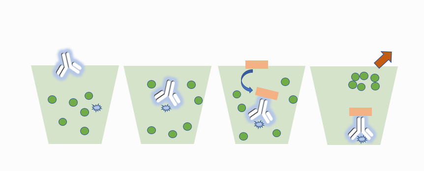
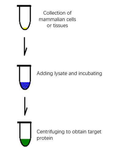
The basic process of protein extraction


Characteristics and Advantages of Abbkine
Protein Extraction Kit:


  • High purity and no pollution-the extracted protein has high purity and maintains natural activity, reducing cross contamination;
  • Strong timeliness-non-denatured active protein can be purified within two hours;
  • Compatible application-the extracted protein can be directly used in subsequent proteomics applications;
  • Follow-up worry-free-kits own a special protease inhibitor package, anti-degradation, anti-useless;
  • High cost performance.

Characteristics and Advantages of Abbkine Protease Inhibitor
Cocktail (100X):


Abbkine Protease Inhibitor Cocktail is an
universal protease inhibitor cocktail contains individual components, including
AEBSF, Aprotinin, Bestatin, E-64, Leupeptin and Pepstatin A with a broad
specificity for cysteine, serine, acid proteases, and aminopeptidases. This
protease inhibitor cocktail has been optimized and tested for mammalian cells
and tissue extracts.


You deserve to have good sample preparation
products!

2019年12月23日星期一

Abbkine Holiday Arrangement of 2020 Spring Festival

Dear partners:


Thank you all for giving our
company full support in 2019. With the approaching of 2020 Spring Festival,
here Abbkine makes the following holiday arrangement:


Holiday Time: 20th
Jan. 2020-31th Jan. 2020 (12 Days).


Holiday Time for the
logistics department: 18th Jan. 2020-2nd Feb. 2020 (16
Days).


Sorry for the inconvenience
caused by the holiday. My sincere regards.


Abbkine Scientific Inc


23th Dec.2019

2019年12月19日星期四

Selected primary antibody| GSK3β Mouse Monoclonal antibody recommendation

Review of Related Articles:


Research
Hotspots of Cell Apoptosis: Bcl-2 Monoclonal Antibody


Abbkine
P53 antibody helps basic cancer research!


Marker
of myofibroblast antibody-α-SMA/ACTA2 Monoclonal Antibody


Selected
primary antibody|TNF-α antibody recommendation


Glycogen synthase kinase-3 (GSK-3) is a
serine-threonine kinase that has been evolutionarily very conservative.The
protein encoded by GSK3β (glycogen synthase kinase 3 beta) is a
serine-threonine kinase, belonging to the glycogen synthase kinase subfamily.
It is involved in energy metabolism, neuronal cell development, and body
pattern formation.


GSK-3β can also act on many signaling
protein structural proteins and transcription factors, and regulate cell
differentiation, proliferation, survival and apoptosis. GSK-3β is a key enzyme
involved in liver glucose metabolism. It inhibits its activity by
phosphorylating GS, reduces liver glucose synthesis, increases blood glucose
concentration in the body, is controlled by insulin in the insulin signaling
pathway, and participates in liver glucose metabolism.


Polymorphisms in GSK3β have been implicated in modifying risk of Parkinson disease, and studies in mice show that overexpression of GSK3β may be relevant to the pathogenesis of Alzheimer disease. Alternatively spliced transcript variants encoding different isoforms have been found for GSK3β .


The picture is from https://link.springer.com/article/10.1007/s12035-013-8571-y


AbbkineGSK3β Mouse Monoclonal antibody (1A6) helps basic research! Basic information about the product:


Product name

#CAT

Reactivity

Applications

Dilution ratio

Observed Band(KD)

GSK3β Mouse Monoclonal antibody (1A6)

ABM0076
human/mouse/rat
WB/IHC-P
WB (1:1000-1:2000) IHC-P (1:100-1:200)
46 kD

GSK3β Mouse Monoclonal antibody (1A6)
verification results:


Immunohistochemical analysis of paraffin-embedded Human Breast Carcinoma Tissue using GSK3β Mouse mAb diluted at 1:200.

Immunohistochemical analysis of paraffin-embedded Human Stomach Carcinoma Tissue using GSK3β Mouse mAb diluted at 1:200.

Western blot analysis of 1) Hela Cell Lysate, 2) 3T3 Cell Lysate, 3) Rat Brain Tissue Lysate using GSK3β Mouse mAb diluted at 1:1000.

2019年12月11日星期三

Selected primary antibody| Cleaved Caspase-3 antibody recommendation

Review of Related Articles:


Research
Hotspots of Cell Apoptosis: Bcl-2 Monoclonal Antibody


Abbkine
P53 antibody helps basic cancer research!


Marker
of myofibroblast antibody-α-SMA/ACTA2 Monoclonal Antibody


Selected
primary antibody|TNF-α antibody recommendation


Cleaved Caspase-3 is an Activated Form of
Caspase-3. Caspases are a family of cysteine proteases that are key mediators
of programmed cell death or apoptosis. The precursor form of all caspases is
composed of a prodomain, and large and small catalytic subunits. The active
forms of caspases are generated by several stimuli including ligand-receptor
interactions, growth factor deprivation and inhibitors of cellular functions.


All known caspases require cleavage
adjacent to aspartates to liberate one large and one small subunit, which
associate into a2b2 tetramer to form the active enzyme. Gene for Caspase-3 also
known as Yama, CPP32, and apopain codes for a 32-kDa protein. The main
substrate of Caspase-3 is poly (ADP- ribose) polymerase PARP (ADP-ribose) which
is related to DNA repair and gene integrity monitoring. When apoptosis is
initiated, Caspase-3 is activated by cleavage of Asp-28/Ser-29 (between
N-terminal prodomains) and Asp-175/Ser-176 (between large subunit and small
subunit), resulting in a large subunit of 17kDa and a small subunit of 12kDa.
P17 and p12 dimerize to form cleaved caspase-3 in activated form.


The picture is from https://www.nature.com/articles/cdd2009180.


AbbkineCleaved-Caspase-3
p17 (D175) Polyclonal Antibody helps basic cancer research! Basic information
about the product:


Product name

#CAT

Reactivity

Applications

Dilution ratio

Observed Band(KD)

Cleaved-Caspase-3 p17 (D175) Polyclonal
Antibody

ABP0021
human/mouse/rat
WB/IF/IHC-P/ELISA

WB (1:500-1:2000)
IF (1:50-1:300)
IHC-P (1:50-1:300)
ELISA(1:20000)

19/17 kD

Abbkine Cleaved-Caspase-3 p17 (D175)
Polyclonal Antibody verification results:


Western Blot analysis using Abbkine Cleaved-Caspase-3 p17 (D175) Polyclonal Antibody.


Immunohistochemical analysis of
paraffin-embedded mouse colon tissue (1:200) using Abbkine Cleaved-Caspase-3
p17 (D175) Polyclonal Antibody. (1:200).

Immunohistochemical analysis of
paraffin-embedded mouse colon tissue (1:200) using Abbkine Cleaved-Caspase-3
p17 (D175) Polyclonal Antibody. (1:200).

2019年11月27日星期三

Selected primary antibody|TNF-α antibody recommendation

Review of Related Articles:


Research
Hotspots of Cell Apoptosis: Bcl-2 Monoclonal Antibody


Abbkine
P53 antibody helps basic cancer research!


Marker of myofibroblast antibody-α-SMA/ACTA2 Monoclonal Antibody


Tumor necrosis factor (TNF, cachexin, or
cachectin; once named as tumor necrosis factor alpha or TNFα) is a cell
signaling protein (cytokine) involved in systemic inflammation and is one of
the cytokines that make up the acute phase reaction. It is produced chiefly by
activated macrophages, although it can be produced by many other cell types
such as CD4+ lymphocytes, NK cells, neutrophils, mast cells, eosinophils, and
neurons. TNF is a member of the TNF superfamily, consisting of various
transmembrane proteins with a homologous TNF domain.


The primary role of TNF is in the regulation of immune cells. TNF, being an endogenous pyrogen, is able to induce fever, apoptotic cell death, cachexia, inflammation and to inhibit tumorigenesis and viral replication and respond to sepsis via IL1- & IL6-producing cells. Dysregulation of TNF production has been implicated in a variety of human diseases including Alzheimer's disease, cancer, major depression, psoriasis and inflammatory bowel disease (IBD). Though controversial, studies of depression and IBD are currently being linked to increased levels of TNF. Recombinant TNF is used as an immunostimulant under the INN tasonermin. TNF can be produced ectopically in the setting of malignancy and parallels parathyroid hormone both in causing secondary hypercalcemia and in the cancers with which excessive production is associated.


Fig: Anti-TNF-α therapies: the next generation (https://www.nature.com/articles/nrd1175)

Abbkine TNF-α
Polyclonal Antibody
helps basic cancer research! Basic information about
the product:


Product name

#CAT

Reactivity

Applications

Dilution ratio

Observed Band(KD)

TNF-α Polyclonal Antibody

ABP0127

Human/Mouse/Rat

WB/IF/IHC-P/ELISA

WB (1:500-1:2000)
IF (1:200-1:1000)
IHC-P (1:100-1:300)
ELISA(1/10000)

16KD

Abbkine TNF-α Polyclonal Antibody verification results:


Western Blot analysis using Abbkine TNF-α Polyclonal Antibody.

Immunofluorescence analysis of human
stomach tissue using Abbkine TNF-α Polyclonal Antibody (1:200).

Immunohistochemical analysis of
paraffin-embedded Human stomach (1:100).

2019年11月14日星期四

Marker of myofibroblast antibody-α-SMA/ACTA2 Monoclonal Antibody

ACTA2 (actin alpha 2) is an actin protein
with several aliases including alpha-actin, alpha-actin-2, aortic smooth muscle
or alpha smooth muscle actin (α-SMA, SMactin, alpha-SM-actin, ASMA). Actins are
a family of globular multi-functional proteins that form microfilaments. ACTA2
is one of 6 different actin isoforms and is involved in the contractile
apparatus of smooth muscle. ACTA2 (as with all the actins) is extremely highly
conserved and found in nearly all mammals.


In humans, ACTA2 is encoded by the ACTA2
gene located on 10q22-q24. Mutations in this gene cause a variety of vascular
diseases, such as thoracic aortic disease, coronary artery disease, stroke,
Moyamoya disease, and multisystemic smooth muscle dysfunction syndrome.


ACTA2 (commonly referred to as alpha-smooth muscle actin or α-SMA) is often used as a marker of myofibroblast formation.


Figure: Diverse origins of the
myofibroblast—implications for kidney fibrosis (From Nature Reviews)


Abbkine α-SMA Monoclonal Antibody Helps Basic Cancer Research! Basic information about the product:


Product name

#CAT

Reactivity

Applications

Dilution ratio

Observed Band(KD)

α-SMA Monoclonal
Antibody


ABM0052


Human/Mouse/ Rat

WB/IF/IHC-P

WB (1:10000-1:100000)
IF (1:100-1:200)
IHC-P (1:1000-1:2000)

42KD


Abbkine α-SMA Monoclonal Antibody verification results:


α-SMA-IF.png

Western blot analysis of 1) Hela, 2) 3T3,
3) rat brain using α-SMA Monoclonal Antibody.
α-SMA-IHC

Immunofluorescence analysis of human
liver tissue.


α-SMA-WB

Immunohistochemical analysis of paraffin-embedded
mouse liver tissue

2019年11月4日星期一

Abbkine winter promotion: looking for you who need live-cell probe

Promotion time: From now to 15th January, 2020.


Promotion product
list:
TraKine™ Pro product
line (Tubulin, Lysosome, Mitochondrion, Nuclei live-cell probe ).


Details of activities: 20% off for TraKine™ Pro product line.


Details for promotion product:


Product name

#Cat

Target

Ex/Em (nm)

Size

Promotion price ($)

TraKine™ Pro Live-cell Tubulin Staining
kit (Green Fluorescence with Super Resolution)

KTC4100

Tubulin

500/520

50/250T

144/496

TraKine™ Pro Live-cell Lysosome Staining
kit (Deep Red Fluorescence with Super Resolution)

KTC4210

Lysosome

650/665

50/250T

128/472

TraKine™ Pro Live-cell Lysosome Staining
kit (Orange Fluorescence with Super Resolution)

KTC4220

Lysosome

565/590

50/250T

128/4721

TraKine™ Pro Live-cell Mitochondrion
Staining kit (Deep Red Fluorescence with Super Resolution)

KTC4300

Mitochondrion

647/661

50/250T

176/576

TraKine™ Pro Live-cell Nuclei Staining
kit (Deep Red Fluorescence with Super Resolution)

KTC4510

Nuclei

650/665

50/250T

144/496


TraKine™ Pro features & benefits:


  • Proprietary probe contains fluorescent dye and a unit which
    electively recognize organelles, with high specificity, low background,
    excellent photostability and good cell permeability.
  • Optimized staining protocol for labeling subcellular structures in
    mammalian living and fixed cells.
  • Fuorescence can last for several hours in cells, stable and
    persistent, especially ideal for monitoring dynamics.
  • Super resolution, especially suitable for Confocal and long-term
    super-resolution imaging (such as SIM, STED, TIRF, STORM and PALM).
  • Safe, very low or no cytotoxicity to cells.

2019年10月30日星期三

Abbkine P53 antibody helps basic cancer research!

P53 gene, human tumor suppressor gene. The gene encodes a protein with a molecular weight of 43.7KDa, but is named P53 because the protein band appears at 53KDa shown by Marker. Wild-type p53 causes cancer cells to apoptosis, thus preventing canceration; But also has the function of helping cell genes repair defects; Studies have found that mutant p53 can enhance canceration. 50% of human tumors are related to P53 gene. Currently, there are liver cancer, breast cancer, bladder cancer, gastric cancer, colon cancer, prostate cancer, soft tissue sarcoma, ovarian cancer, brain tumor, lymphocyte tumor, esophageal cancer, lung cancer, osteosarcoma, etc. P53 mutation in human tumors is mainly in highly conserved regions, with the highest mutation at 175, 248, 249, 273, 282 sites, and different types of tumors.  


P53 has a very short half-life, so it is almost undetectable in unstressed cells. This is mainly realized by the continuous ubiquitination of p53 and the degradation of 26S proteasome in the later stage under the action of its inhibitor MDM2. Cell stress signals such as DNA damage can inhibit ubiquitination of p53 and lead to its stabilization and activation. Once p53 is activated, it will bind to its downstream target regulatory elements and regulate its transcription. Therefore, various cell therapy schemes based on different tumor inhibition functions of p53 can be started. Just because past studies have shown that p53 gene mutations exist in about half of cancers, scientists regard them as important targets for developing new cancer therapies.


Mutant p53 promotes adaptive responses to cancer-related stress conditions to support tumor progression.

Abbkine P53 Antibody Helps Basic Cancer
Research! Basic information about the product:


Product name
#CAT
Reactivity Applications
Dilution ratio
p53 Polyclonal Antibody ABP0110   Human, Monkey, Mouse, Rat
WB/IF/IHC-P/ELISA

WB (1:500-1:2000)
IF (1:200-1:1000)
IHC-P (1:100-1:300)
ELISA  (1:10000).

Abbkine P53 Antibody verification results:


Western Blot analysis of various cells using p53 Polyclonal Antibody diluted at 1:1000.
Immunofluorescence analysis of human liver tissue. 1, p53 Polyclonal Antibody (red) was diluted at 1:200 (4°C, overnight). 2, Cy3 Labeled secondary antibody was diluted at 1:300 (room temperature, 50min). 3, Picture B: DAPI (blue) 10min. Picture A: Target. Picture B: DAPI. Picture C: merge of A+B.
Immunohistochemical analysis of paraffin-embedded mouse testis tissue. 1, p53 Polyclonal Antibody was diluted at 1:200 (4°C, overnight). 2, Sodium citrate pH 6.0 was used for antibody retrieval (>98°C, 20min). 3, secondary antibody was diluted at 1:200 (room temperature, 30min). Negative control was used by secondary antibody only.

2019年10月15日星期二

IL-6: Target Molecule for Cytokine Storms

Interleukin-6 (IL-6) is a multi-effect cytokine
that plays an important role in host defense by regulating immune and
inflammatory reactions. IL-6 is mainly produced by macrophages, T lymphocytes
and B lymphocytes. When the body has inflammatory reaction or infection,
various cytokines such as virus, endotoxin and tumor necrosis factor can induce
the body to produce IL-6. The biological effects of IL-6 are mainly reflected
in inducing B cell differentiation to produce immunoglobulin, promoting T cell
proliferation and growth, enhancing blood cell differentiation and its
anti-tumor effect, promoting bone marrow hematopoietic stem cell proliferation,
etc.


IL-6 is an important member of cytokine network and plays a central role in acute inflammatory reaction. The production of IL-6 induces the production of C-reactive protein (CRP) and procalcitonin (PCT), which are directly related to inflammatory diseases and infection degree. IL-6 can diagnose early inflammation and warn the occurrence of sepsis faster. In addition, the half-life of IL-6 is shorter than CRP and PCT, which can reflect the effect of antibiotic treatment faster and better reflect the prognosis of patients. At the same time, IL-6 can be used as a biomarker of disease severity and prognosis indicators in cytokine storms, and its expression is superior to TNF-α and IL-1. A large number of research results show that IL-6 is a suitable target molecule for cytokine storms.


If you are studying IL-6 or are about to study IL-6, the recommended IL-6 cytokines are as follows:


Product
name


#cat

Sequence

Purity

Expressed
Host


Endotoxin

Formulation

Human
IL-6 protein

PRP100120

Human IL6
(NP_000591.1) (Val 30-Met 212)

>
95%

E.
coli

<1EU/μg

lyophilized
powder

Mouse IL-6 protein

PRP100371

Mouse
IL6 (P08505) (Phe25-Thr211)

>
95%

E.
coli

<1EU/μg

lyophilized
powder

SDS-PAGE analysis:


Human IL-6 protein


Mouse IL-6 protein


Recommendations
for cytokine storage:


Cytokines
in lyophilized powder form are very stable and can be stored for several years
at -20℃ or -80℃. Freeze-dried powder should be dissolved before use and then
added to the culture medium in liquid form. The dissolved cytokines can only be
stored for a short time (about 1 week) at 4℃. For long-term preservation, first
prepare a diluent (which must contain carrier protein, such as 0.1% BSA, 5%HSA,
or 10% FBS), and then sub-pack and freeze at -20℃ or -80℃. Repeated freezing
and thawing must be avoided, as each freezing and thawing will cause partial
inactivation of proteins.

2019年10月10日星期四

Abbkine Meets You at 17th International Immunology Congress (IUIS 2019)

Abbkine
invites you to visit IUIS 2019, Beijing, China.


Abbkine featured products and services will be displayed at the exhibition. There always be one type can flash into your eyes. Participate in interaction, more surprises are waiting for you!


Date: October
19 – 23, 2019


Booth:#D22,level 4


Address: China
National Convention Center, Beijing, China


About: The 17th International Congress of Immunology (IUIS 2019) will be held from October 19 – 23, 2019 at the China National Convention Center, Beijing, China. IUIS 2019 promises to be an unforgettable event that will bring together delegates from all over the world. We anticipate over 5000 participants, including international leaders at the forefront of the discipline that will present the most recent advances in basic immunology and clinical translation.


2019年9月24日星期二

In 4 years, Abbkine helped more than 1200 English articles to be published. Impact factors exceed 4500 points! ! !

Abbkine focuses on immunology and cytology, and is committed to innovating and developing various antibodies, proteins, analytical reagents and kits, with a view to becoming a key promoter in the fields of life science research and development, drug research and development, etc.


Some articles published using Abbkine products:


1. The negative effect of silica nanoparticles on adipogenic differentiation of human mesenchymal stem cells. Materials Science and Engineering. (IF: 27.24)


Using Abbkine Dylight 594, Goat Anti-Mouse IgG


2. Bph6 encodes an exocyst-localized protein and confers broad resistance to planthoppers in rice. NATURE GENETICS. (IF: 19.88)


Using Abbkine Anti-Plant Actin Mouse Monoclonal Antibody (3T3)


3. The deubiquitinating enzyme cylindromatosis mitigates nonalcoholic steatohepatitis. . (IF: 19.14)


Using Abbkine HRP, Goat Anti-Rabbit IgG


4. Research on the interaction of protein Tmub1 and securin after partial hepatectomy. Journal of the American Chemical Society. (IF: 14.75)


Using Abbkine IFKine™ Red Donkey Anti-Rabbit IgG


5. Verification of Differential Expression Genes after CacyBP/SIP Nuclear Translocation in Colon Carcinoma Cell Line. Journal of the American Chemical Society. (IF: 14.75)


Using Abbkine Anti-PCNA Mouse Monoclonal Antibody (1D7)


6. CD8+T cells induce cachexia during chronic viral infection. NATURE IMMUNOLOGY. (IF: 14.71)


Using Abbkine EliKine™ Free Triiodothyronine (fT3) ELISA Kit


7. SET8 prevents excessive DNA methylation by methylation-mediated degradation of UHRF1 and DNMT1. Nucleic Acids Research. (IF: 11.14)


Using Abbkine Pan Methylated Lysine Monoclonal Antibody (Mix)


8. The lncRNA PVT1 regulates nasopharyngeal carcinoma cell proliferation via activating the KAT2A acetyltransferase and stabilizing HIF-1α. Cell Death & Differentiation. (IF: 8.086)


Using Abbkine KAT6A
Polyclonal Antibody


9. DIPK2A promotes STX17- and VAMP7-mediated autophagosome-lysosome fusion by binding to VAMP7B. Autophagy. (IF: 7.01)


Using Abbkine IPKine™
HRP, Goat Anti-Mouse IgG HCS


10. CAV1-CAVIN1-LC3B-mediated autophagy regulates high glucose-stimulated LDL transcytosis. Autophagy. (IF: 7.01)


Using Abbkine IPKine™ HRP, Goat Anti-Rabbit IgG HCS


Some partners of
Abbkine:


Harvard University, Georgia Institute of Technology, Oxford University, Duke University, Stanford University, Peking University, Tsinghua University, Zhejiang University, Chinese Academy of Sciences, Wuhan University, Fudan University.


2019年9月10日星期二

Research Hotspots of Cell Apoptosis: Bcl-2 Monoclonal Antibody

【Promotion】Abbkine
Selected Primary Antibody-Click for Promotion
details.


Bcl-2 gene (B-cell lymphoma-2) is a member of Bcl-2 family, which is the earliest discovered apoptosis protein. It is an anti-apoptosis protein containing multiple BH domains. It is highly expressed in some cancer cells and selectively plays an anti-tumor role. Bcl-2 family proteins play an important role in the regulation of apoptosis by forming dimers with Bax and dimerizing themselves. When Bcl-2 protein is inhibited, its dimer with Bax decreases, leading to cell apoptosis. When Bcl-2 protein is overexpressed, heterodimers formed by Bcl-2 protein and Bax increase and apoptosis is inhibited. The balance between Bcl-2 and Bax proteins at the cell death signal checkpoint determines the survival or apoptosis of cells.


Fig: Bcl-2 and Cell apoptosis

We recommend Abbkine selected Bcl-2 antibody for WB, IHC-P and IF experiments in human, rats, mice and chickens.


Product name

Cat#

Application

MW

Bcl-2 Monoclonal Antibody

ABM0010

WB(1:1000-2000),IF(1:50-200),IHC-P(1:200)

26KD


The result:


1) Hela cells; 2) MCF-7 cells; Using Abbkine Bcl-2 monoclonal antibody (ABM0010), WB 1:1000 dilution.


Human oviduct, Using Abbkine Bcl-2 monoclonal antibody (ABM0010) and IHC-P 1:100.


NIH/3T3 cells, Using Abbkine Bcl-2 monoclonal antibody (ABM0010), green, IF 1:100.

2019年9月2日星期一

Beyond BrdU, New Generation Cell Proliferation Imaging Analysis Kit (EdU Method)

Common methods of cell proliferation
mainly include MTT method, WST-1 method and CCK-8 method, which are mainly
based on the activity of cells, thus reflecting the overall proliferation
effect, but unable to detect individual proliferating cells. Recognized as the
most accurate method to detect cell proliferation is to directly detect DNA
synthesis in cells. Previously, BrdU method was the most commonly used method.
However, the method requires DNA denaturation (such as acid denaturation,
thermal denaturation or DNase digestion) to expose BrdU, thus binding to BrdU
antibody. The whole experiment has many influencing factors and poor stability.


Abbkine innovates and develops EdU
method based on EdU incorporation and subsequent click reaction to solve this
application difficulty:


Product name

Cat#

Fluorescent characteristic

Cell Proliferation EdU Image Kit
(Green Fluorescence)

KTA2030

AbFluor
488
Ex/Em = 501/525 nm

Cell Proliferation EdU Image Kit
(Orange Fluorescence)

KTA2031

AbFluor
545
Ex/Em = 546/565 nm


Principle:EdU (5-ethynyl-2´-deoxyuridine) is a nucleoside analog of thymidine and is incorporated into DNA during active DNA synthesis. Comparing to BrdU assays, the EdU-Click Assays are not antibody based and therefore do not require DNA denaturation (typically using HCl or heat or digestion with DNase) for detection of the incorporated nucleoside. Detection is based on a click reaction, a copper-catalyzed covalent reaction between an azide and an alkyne, is complete within 30 minutes.


Kit components

Advantage
• EdU (10mM)
• AbFluor 488 azide or AbFluor 545 azide
• 10×Reaction buffer
• Copper
• Reducing Agent
• Not antibody based 
• No DNA denaturation. Can maintain cell morphology and DNA integrity
• Patented AbFluor 488 and 545 azide have good light stability and quenching resistance
• Optimization for fluorescence microscope


Results:


KTA2030
Imaging: Hela Cells (Green)

KTA2031
Imaging: Hela Cells (Red)

2019年8月26日星期一

Loading Control & Tag Antibody-Buy two get one for free

Flash sale: From now to October 31st, 2019


Promotion product list: Classical loading control and tag antibodies and the conjugated versions of these antibodies, including HRP, Biotin, FITC, Cy3, Cy5 and AbFlour TM dyes.


Details of activities: Buy 2 get 1 for free (if a single order is full of 3 antibodies, the antibody with the lowest price will be exempted).


Why choose Abbkine loading control and tag antibodies?


  • Abbkine offers a wide range of
    loading control and tag antibodies, including the hot targets and unique
    species, covering the molecular weights from 15 kDa to 116 kDa.
  • Abbkine conjugated loading
    control and tag antibodies have diverse conjugates, including enzyme and
    featured fluorescent dyes, with the application of WB, IHC, IF and IP
    experiments.
  • Abbkine loading control and
    tag antibodies are verified through rigorous procedures, popular with customers
    for their good performance and high sensitivity.
  • Different sizes for clients to
    choose, small size and bulk size are available, both with high
    cost-effectiveness.

Part of promotion product list:


Classical loading control and tag antibodies (Citation of over 100 SCI Documents)


Cat#

Product name

Application

size

A01010

Anti-β-Actin
Mouse Monoclonal Antibody (1C7)

WB#IHC-P#IF

50/200/200x5ul

A01020

Anti-GAPDH
Mouse Monoclonal Antibody (2B5)

WB#IHC-P#IF

50/200/200x5ul

A01030

Anti-β-Tubulin
Mouse Monoclonal Antibody (3G6)

WB#IHC-P#IF

50/200/200x5ul

A01050

Anti-Plant
Actin Mouse Monoclonal Antibody (3T3)

WB

50/200/200x5ul

A02010

Anti-DDDDK
Tag Mouse Monoclonal Antibody (1B10)

WB#IF#IP

50/200/200x5ul

A02050

Anti-His
Tag Mouse Monoclonal Antibody (5C3)

WB#IF#IP

50/200/200x5ul

A02040

Anti-HA
Tag Mouse Monoclonal Antibody (4F6)

WB#IF#IP

50/200/200x5ul

A02020

Anti-GFP
Tag Mouse Monoclonal Antibody (3D3)

WB#IP

50/200/200x5ul

A02030

Anti-GST
Tag Mouse Monoclonal Antibody (2A8)

WB

50/200/200x5ul

A02060

Anti-Myc
Tag Mouse Monoclonal Antibody (2D5)

WB#IF#IP

50/200/200x5ul

A02080

Anti-mCherry
Tag Mouse Monoclonal Antibody (9D3)

WB#IF

50/200/200x5ul


IP experiment partner-Agarose/Magnetic beads conjugated tag antibodies


Compared
with the traditional immunoprecipitation (IP) method using Protein A/G,
Abbkine's agarose/magnetic bead coupling labeled antibody eliminates the step
of Protein A/G binding with antigen-antibody complex. After the conjugated
antibody is directly combined with the target protein, the target protein is
separated from the cell lysate by centrifugation or using a magnetic rack. It
not only makes the experiment simple and fast, but also can avoid non-specific
binding and reduce the background.


Cat#

Product name

Application

size

A02010AGB

Anti-DDDDK
Tag Mouse Mab (1B10), Agarose

IP

100ul/400ul/2ml

A02010MGB

Anti-DDDDK
Tag Mouse Mab (1B10), Magnetic Beads

IP

100ul/400ul/2ml

A02040AGB

Anti-HA
Tag Mouse Mab (4F6), Agarose

IP

100ul/400ul/2ml

A02040MGB

Anti-HA
Tag Mouse Mab (4F6), Magnetic Beads

IP

100ul/400ul/2ml

A02050AGB

Anti-His
Tag Mouse Mab (5C3), Agarose

IP

100ul/400ul/2ml

A02050MGB

Anti-His
Tag Mouse Mab (5C3), Magnetic Beads

IP

100ul/400ul/2ml

A02060AGB

Anti-Myc
Tag Mouse Mab (2D5), Agarose

IP

100ul/400ul/2ml

A02060MGB

Anti-Myc
Tag Mouse Mab (2D5), Magnetic Beads

IP

100ul/400ul/2ml

A02170AGB

Anti-V5
Tag Mouse Mab (11D5), Agarose

IP

100ul/400ul/2ml

A02170MGB

Anti-V5
Tag Mouse Mab (11D5), Magnetic Beads

IP

100ul/400ul/2ml

A02180AGB

Anti-VSV-G-Tag
Mouse Mab (14D2), Agarose

IP

100ul/400ul/2ml

A02180MGB

Anti-VSV-G-Tag
Mouse Mab (14D2), Magnetic Beads

IP

100ul/400ul/2ml

For
the full product promotion list, please contact Abbkine's local distributor or
contact service@abbkine.com directly. Abbkine has the final right to interpret
this activity.

2019年8月23日星期五

Selection Strategy of Apoptosis Products- Late apoptosis

Apoptosis
is an orderly and programmed death followed by cells under the influence of
physiological or pathological factors in order to maintain internal environment
stability. Apoptosis occurs in cells. First, the cell volume shrinks and the
connection disappears. Then, the density of cytoplasm increases, mitochondrial
membrane potential disappears, permeability changes, cytochrome C is released
to cytoplasm, nuclear substance is concentrated, nuclear membrane nucleolus is broken,
DNA is degraded into fragments, and finally apoptotic bodies are formed and
engulfed by macrophages.


Late apoptosis


In cell apoptosis, especially in the late stage of apoptosis, chromosomal
DNA will be broken, resulting in a large number of sticky 3'-OH ends. Under the
action of deoxynucleotide terminal transferase (TdT), derivatives formed by
deoxyuridine triphosphate nucleotide (dUTP) and fluorescein can be labeled to
the 3'- end of DNA, namely deoxynucleotide terminal transferase mediated nick
end labeling (TUNEL). Normal or proliferating cells have few DNA breaks and can
rarely be stained. Therefore, TUNEL is the most commonly used method to detect
DNA fragmentation in late apoptosis.


Product recommendation


Product name

Cat#

TUNEL Apoptosis Detection Kit (Green
Fluorescence)

KTA2010

TUNEL Apoptosis Detection Kit (Orange
Fluorescence)

KTA2011


Product character


Abbkine's TUNEL apoptosis detection kit provides a complete set of
reagent components and optimized experimental scheme required for the
experiment. It is suitable for a variety of instruments such as fluorescence
enzyme labeling instrument, fluorescence microscope, flow cytometer, etc. It
can be used to detect adherent cells, suspended cells, paraffin-embedded tissue
sections, frozen sections and other sample types.


Apoptosis cannot be stopped once it
starts, it is a highly regulated process.


Apoptosis can be
initiated by two initial pathways. The internal initial pathway is activated by
non-receptor stimulation, such as DNA damage, endoplasmic reticulum stress,
metabolic stress, mitochondrial outer membrane permeability changes, such as
cytochrome C release. The external pathway starts by binding death receptor to
ligand (such as FasL, tumor necrosis factor TNF). After that, the two
approaches converge in caspase cascade reaction, and cell death is induced by
activating caspase protease or protein degrading enzyme. Anti-apoptotic
ligands, such as cytokines and growth factors, promote cell survival,
proliferation and differentiation through different signaling molecules such as
AKT and p90RSK and anti-apoptotic proteins such as Bcl-2 and Bcl-x. The withdrawal
of these cytokines and growth factors leads to cell death.


Product recommendation


Product name

Cat#

Application

Bax Mouse Monoclonal Antibody (6F11)

ABM40273

IF, IHC-p, WB

Bcl-2 Monoclonal Antibody

ABM0010

IF, IHC-p, WB

Bad Polyclonal Antibody

ABP55880

ELISA, IF, IHC-p, WB

p53 Monoclonal Antibody

ABM0016

IF, IHC-p, WB

FAS Polyclonal Antibody

ABP51327

ELISA, IF, IHC-p, WB

Caspase-3 Polyclonal Antibody

ABP52935

ELISA, IF, IHC-p, WB

Caspase-8
Monoclonal Antibody

ABM0053

IF, IHC-p, WB

Caspase 9 Monoclonal Antibody

ABM0028

IF, IHC-p, IP, WB

NFkB p65 Monoclonal Antibody

ABM40111

IF, IHC-p, IP, WB


Product character


Abbkine has selected the antibody of apoptosis series. After multiple strict verifications, it is suitable for a variety of cell applications and meets the research needs of most customers. The excellent results are as follows:


IHC analysis of paraffin-embedded human uterine tissue was performed using Bad polyclonal antibody diluted at 1: 200.


IF analysis of rat lung tissue was performed using Caspase-3 polyclonal antibody diluted at 1: 200.


2019年8月21日星期三

Selection Strategy of Apoptosis Products- Early apoptosis

Apoptosis
is a dynamic process, which involves a series of complex biochemical reactions,
expression regulation of multiple genes, signal transduction, cascade reactions
involving multiple enzymes and multiple signal pathways. The cell feels the
corresponding apoptosis signal stimulation, and a series of control switches in
the cell are turned on or off, and activation of various enzymes triggers a
series of cascade reactions. Different external factors initiate apoptosis in
different ways, resulting in different signal transduction.


Early apoptosis


Feature 1: eversion of lipid membrane
inside


In normal cells, phosphatidylserine (PS) is only distributed on the inner
side of the lipid bilayer of the cell membrane. In the early stage of cell
apoptosis, PS turns from the inner side of the lipid membrane to the outer
side. Annexin V is a calcium-dependent phospholipid binding protein with a
molecular weight of 35-36kD, and has high affinity with PS. It binds to the
cell membrane of early apoptotic cells through PS exposed on the outside of
cells. Therefore Annexin V is a sensitive indicator to detect early apoptosis
of cells. Usually, Annexin V is labeled with some fluorescent probes, which can
simply and directly detect PS eversion, an important feature of apoptosis.


Product recommendation


Product name

Cat#

Abbkine Annexin
V-AbFluor™ 405 Apoptosis Detection Kit

KTA0001

Abbkine Annexin
V-AbFluor™ 488 Apoptosis Detection Kit

KTA0002

Abbkine Annexin
V-AbFluor™ 555 Apoptosis Detection Kit

KTA0003

Abbkine Annexin
V-AbFluor™ 647 Apoptosis Detection Kit

KTA0004


Product character


AbFlourTM
series of characteristic fluorescent dyes (blue, green and red) patented by
Abbkine label recombinant human Annexin V protein, and are matched with PI to
introduce different fluorescence apoptosis detection kits, which are suitable
for detection with fluorescence equipment such as flow cytometry and
fluorescence microscope.


Feature 2: Mitochondrial membrane
potential decreased


Normal mitochondrial membrane potential is the premise for maintaining
mitochondrial oxidative phosphorylation and ATP production, and is necessary
for maintaining mitochondrial function. The decline of mitochondrial membrane
potential is a landmark event in the early stage of cell apoptosis. JC-1 is an
ideal fluorescent probe widely used for detecting mitochondrial membrane
potential. When mitochondrial membrane potential is high, JC-1 aggregates in
the matrix of mitochondria to form polymers (J-aggregates). Red fluorescence
can be generated(Ex/Em =
585/590). when the mitochondrial membrane potential is low, JC-1 cannot
aggregate in the matrix of mitochondria. at this time, JC-1 is a monomer, which
can generate green fluorescence (Ex/Em = 510/527 nm).


Product recommendation


Product name

Cat#

Mitochondrial Membrane Potential Assay Kit
(JC-1)

KTA4001


Product character


Abbkine Mitochondrial Membrane Potential Analysis Kit (JC-1) uses JC-1 as a fluorescent probe to rapidly and sensitively detect changes in mitochondrial membrane potential. This kit can very conveniently detect changes in mitochondrial membrane potential through changes in fluorescence color.


Abbkine Mitochondrial Membrane Potential Analysis Kit (JC-1)

2019年8月7日星期三

Complete solution of immunoprecipitation (IP)-break down the key steps of IP step by step!

Step 1: Understand what immunoprecipitation is? Why should I do
immunoprecipitation?


Immunoprecipitation (IP) is a method for purifying and enriching target proteins from specific mixtures (usually cell lysates or expression supernatants) by using antigen-antibody specific reactions. The traditional IP experiment is that after the antibody is combined with the target protein, it is incubated with agarose or magnetic beads coupled with Protein A /G (Fc fragment of binding antibody), and the bead-protein A/G- antibody-target protein complex is obtained by centrifugation. The complex is washed and eluted for subsequent downstream experiments. IP is an important step in many protein-related researches. It is used to study the existence, relative abundance, up-down regulation of protein expression, protein-to-stability and interaction of proteins.


Traditional IP protocol

Summary: The key tools I need to use in the whole
process of IP are,


1) primary antibody: select the primary antibody that can
react with the sample and do IP experiment;


2) Potein APotein G or Protein
A /G
:Potein A, Potein
G and Protein A /G have different binding abilities to immunoglobulins from
different sources and subtypes. how to choose? Please refer to https://www.abbkine.com/affinity-of-protein-a-protein-g-and-protein-ag/


3) Selection Guide:


Cat#

Product name

BMR20500

PurKine™
Protein A Resin

BMR20504

PurKine™
Protein A Resin 4FF

BMR20600

PurKine™
Protein G Resin

BMR20604

PurKine™
Protein G Resin 4FF

BMR20704

PurKine™
Protein A/G Resin 4FF


Step 2: Finding that there is no suitable primary antibody to meet the immunoprecipitation I need to do?


Some target proteins cannot be immunoprecipitated because there is no
corresponding specific antibody. For example, the antigen epitope recognized by
the antibody is shielded by the interaction protein, unable to interact with
the target protein or the antibody selection is inappropriate, the selected
antibody cannot recognize the protein in natural conformation, etc. After an
epitope tag protein is used, the tag antibody against this epitope can be
selected for immunoprecipitation. Manufacturers will introduce some common
label antibodies for IP experiments, involving labels such as flag, HA, His and
Myc to meet the needs of most customers.


In particular, the agarose/magnetic bead coupled labeled antibody can
optimize the immunoprecipitation experiment to the greatest extent and save
time and cost.


1) Principle: The step of Protein A/G binding with antigen-antibody complex is omitted, and the problem of weak binding ability between Protein A/G and antibody is solved. After the conjugated antibody is directly combined with the target protein, the target protein is separated from the cell lysate by centrifugation or using a magnetic rack. The high-specificity monoclonal label antibody can realize high yield and high purity, and the stable and pre-sealed filler and the specific antibody reduce non-specific binding in the immunoprecipitation process.


Agarose/magnetic bead conjugated label antibody is applied to IP

2) Selection Guide:


Cat#

Product name

A02010AGB

Anti-DDDDK Tag
Mouse Monoclonal Antibody (1B10), Agarose

A02010MGB

Anti-DDDDK Tag
Mouse Monoclonal Antibody (1B10), Magnetic Beads

A02040AGB

Anti-HA Tag
Mouse Monoclonal Antibody (4F6), Agarose

A02040MGB

Anti-HA Tag
Mouse Monoclonal Antibody (4F6), Magnetic Beads

A02050AGB

Anti-His Tag
Mouse Monoclonal Antibody (5C3), Agarose

A02050MGB

Anti-His Tag
Mouse Monoclonal Antibody (5C3), Magnetic Beads

A02060AGB

Anti-Myc Tag
Mouse Monoclonal Antibody (2D5), Agarose

A02060MGB

Anti-Myc Tag
Mouse Monoclonal Antibody (2D5), Magnetic Beads


Step 3: How to deal with heavy chain and light chain interference in WB (IP-WB) after immunoprecipitation?


In WB verification after immunoprecipitation experiment, when
conventional Anti-IgG (H+L) enzyme labeled secondary antibody is used, two
bands corresponding to heavy chain (50kDa) and light chain (25kDa) generated
after denaturation of immunoprecipitation primary antibody will usually appear.
If the molecular weight of the detected target protein is near here, it will be
interfered by the two bands. How to avoid interference,


Method 1: Select the secondary antibody that only
reacts with heavy chain or light chain. If the molecular weight of the target
protein is less than 30KD, in order to avoid interference of antibody light
chain, heavy chain specific secondary antibody is selected; If the molecular
weight of the target protein is more than 30KD, in order to avoid interference
of antibody heavy chain, light chain specific secondary antibody is selected;


Selection Guide:


Cat#

Product name

Description

A25012

IPKine™ HRP,
Goat Anti-Mouse IgG LCS

light chain
specific

A25022

IPKine™ HRP,
Mouse Anti-Rabbit IgG LCS

light chain
specific

A25112

IPKine™ HRP, Goat
Anti-Mouse IgG HCS

Heavy chain
specific

A25222

IPKine™ HRP,
Goat Anti-Rabbit IgG HCS

Heavy chain
specific


Method 2: When selecting WB primary antibody, select different species from IP primary antibody to avoid interference from IP primary antibody;


Method 3: Select the WB primary antibody directly coupled with HRP, and omit the
step of secondary antibody.  Immune
precipitation is broken in three steps.


If you have any questions, welcome to contact
us.

2019年7月31日星期三

Quantitative determination of LDH in samples, Abbkine CheKine™ Lactate Dehydrogenase (LDH) Assay Kit will offer you an easier method

Lactate dehydrogenase (LDH) is an oxidoreductase
found in nearly all living cells (animals, plants, and prokaryotes). LDH
catalyzes the conversion of lactate to pyruvate and back, as it converts NAD+
to NADH and back. A dehydrogenase is an enzyme that transfers a hydride from
one molecule to another.


Quantification of Lactate dehydrogenase has clinical
significance because serum levels of certain lactate dehydrogenase isoenzymes
reflect the pathological conditions of specific tissues. When tissue is damaged
by disease, injury, or toxic substances, lactate dehydrogenase is released into
the bloodstream. Because lactate dehydrogenase is a fairly stable enzyme, it
has been widely used to evaluate tissue, cell damage and toxicity.


Abbkine CheKine™ Lactate Dehydrogenase
(LDH) Assay Kit, more simple, convenient and sensitive


Product Name

CAT #

CheKine™ Lactate Dehydrogenase Assay
Kit


KTB1110


【Principle】


provides a simple and easy colorimetric assay for measuring LDH in samples. In this colorimetric assay, LDH reduces NAD to NADH, which then interacts with a probe to produce a color (λmax = 450 nm), the LDH present in the sample is proportional to the signal obtained.


【Samples】


serum, plasma, cell culture supernatants, tissue/cell lysates, culture medium, fermentation and other biological fluids.


【Kit components】


Name

Storage temperature

Function

Assay Buffer
4ºC maintain reaction environment

Lactate
-20ºC LDH substrate

NAD
-20ºC LDH substrate

WST-8
-20ºC, protect from light coloration

Enhancer
-20ºC, protect from light electron transport complex

LDH
Standard

-20ºC Prepare standard curve


【Procedure】


  1. Working Reagent Preparation
  2. Add standard and sample per
    well, then add Working Reagent to each well. Tap plate to mix. Immediately read
    optical density at 450nm (OD0).
  3. Incubate for 30 min at 37°C in the dark.
    Read optical density at 450nm again (OD30).

【Standard Curve】


About Abbkine Scientific Co., Ltd.


Abbkine serves global scientists in the
field of proteomics and cytology and is committed to the innovation and
development of various scientific reagents related to proteomics and cytology,
expecting to accelerate the pace of life science research and drug discovery.
Proteomics products cover the preparation of samples (protein extraction,
purification, coupling), protein quantification, antibodies and kits for
protein detection. Cytology products involve cytokines (cell culture), cell
status detection, cell staining, organelle extraction, cell metabolism and
cytopathology reagents (kits). Abbkine relies on the product portfolio and
unique marketing support as the main market strategy and product innovation
mode, with ultimate aim to facilitate your research career.

2019年7月23日星期二

CheKine™ Xanthine Oxidase Assay Kit, a great tool for study of hyperuricemia


Xanthine oxidase is a form of xanthine oxidoreductase, a type of enzyme that generates reactive oxygen species. These enzymes catalyze the oxidation of hypoxanthine to xanthine and can further catalyze the oxidation of xanthine to uric acid. These enzymes play an important role in the catabolism of purines in some species, including humans.


Xanthine oxidase (XO, EC 1.17.3.2 ) is present in appreciable amounts in the liver and jejunum in healthy individuals. Most of the protein in the liver exists in a form with xanthine dehydrogenase activity, but it can be converted to xanthine oxidase by reversible sulfhydryl oxidation or by irreversible proteolytic modification. In various liver disorders, XO is released into circulation. Therefore, determination of serum XO level serves as a sensitive indicator of acute liver damage such as jaundice.


Abbkine CheKine™ Xanthine Oxidase Assay Kit provides a simple and easy colorimetric assay for the quantitative determination of the XO present in a variety of samples. In the assay, XO oxidizes xanthine to superoxide (O2 - ). Superoxide (O2 - ) reacts with a tetrazolium salt WST-8 dye to form a water-soluble colored formazan product, which can be easily quantified colorimetrically at OD 450 nm. Therefore, the Xanthine Oxidase present in the sample is proportional to the signal obtained.


Product
Name

CAT #

Detection
Range

CheKine™
Xanthine Oxidase Assay Kit

KTB1070

15.6
-500 mU/mL


【Kit components】


Assay Buffer


Sample Diluent


WST-8


Enhancer


Xanthine Oxidase Standard (10 U/mL)


Xanthine


【Procedure】


  1. Prepare
    the Working Reagent.
  2. Add
    standard and sample per well. Then add Working Reagent per well quickly. Tap
    plate to mix briefly and thoroughly. Immediately read
    optical density at 450nm (OD0).
  3. Incubate for 30 min at room
    temperature (25°C) in the dark. Read optical density at 450nm again (OD30).

【Standard Curve】


About Abbkine Scientific Co., Ltd.


Abbkine serves global scientists in the field of
proteomics and cytology and is committed to the innovation and development of
various scientific reagents related to proteomics and cytology, expecting to
accelerate the pace of life science research and drug discovery. Proteomics
products cover the preparation of samples (protein extraction, purification,
coupling), protein quantification, antibodies and kits for protein detection.
Cytology products involve cytokines (cell culture), cell status detection, cell
staining, organelle extraction, cell metabolism and cytopathology reagents
(kits). Abbkine relies on the product portfolio and unique marketing support as
the main market strategy and product innovation mode, with ultimate aim to
facilitate your research career.

2019年7月16日星期二

New biosensor weapons, CheKine™ Glucose Oxidase Activity (GOD)Assay Kit

Glucose oxidase is a an oxido-reductase
that catalyses the oxidation of glucose to hydrogen peroxide and
D-glucono-δ-lactone. This enzyme is produced by certain species of fungi and
insects and displays antibacterial activity when oxygen and glucose are
present. Glucose oxidase is widely used to measure glucose in body fluids, as
well as residual glucose and oxygen from beverages, foods and other
agricultural products. In addition, glucose oxidase is commonly used as
biosensors to detect glucose.


Test the activity of glucose oxidase
(GOD), recommend the Abbkine CheKine™ glucose oxidase (GOD) activity kit for
you, details are as follows:


Product Name

CAT #

Detection Range

CheKine™ Glucose Oxidase Activity
(GOD)Assay Kit

KTB1310

0.05 - 2 U/L


【Principle】


In this assay, GOD activity
is determined by a coupled enzyme assay, in which GOD oxidizes D-glucose
resulting in the production of hydrogen peroxide (H2O2) that reacts with
chromogen in an acid condition, produce a product that can be measured at OD
580 nm. Therefore, the glucose oxidase activity present in the sample is
proportional to the signal obtained.


【Sample】


Serum, Plasma, Cells, Tissues, Urine, Food,
Medium, other body fluids.


Kit components


  • Assay Buffer (20x)
  • Glucose (0.2 M)
  • H2O2 Standard (0.88 M)
  • Chromogen
  • Glucose Oxidase (control)

Simple procedure


  1. Prepare standards and sample, add Glucose
    to each tube, mix and incubate for 20 min at 37°C.
  2. Transfer the mixture into a clear bottom
    96-well plate, add Chromogen per well, mix and read optical density at 580nm(OD0).
    Incubate for 10 min at 37°C in the dark. Read optical density at 580nm again(OD10).

【Standard Curve】


About Abbkine Scientific Co., Ltd.


Abbkine serves global scientists in the
field of proteomics and cytology and is committed to the innovation and
development of various scientific reagents related to proteomics and cytology,
expecting to accelerate the pace of life science research and drug discovery.
Proteomics products cover the preparation of samples (protein extraction,
purification, coupling), protein quantification, antibodies and kits for
protein detection. Cytology products involve cytokines (cell culture), cell
status detection, cell staining, organelle extraction, cell metabolism and
cytopathology reagents (kits). Abbkine relies on the product portfolio and
unique marketing support as the main market strategy and product innovation mode,
with ultimate aim to facilitate your research career.

CheKine™ Lactate Assay Kit, fast quantitative determination of L (+)-Lactate in various samples

Lactate
(CH3CH(OH)COO-) is a metabolic compound formed in animals by the action of the enzyme
lactate dehydrogenase. Lactate is produced in proliferating cells during
anaerobic conditions such as exercise. Abnormally high concentrations of
lactate have been related to pathological conditions such as cancer, diabetes,
and lactate acidosis.


L(+)-Lactate is the major lactate stereoisomer formed in human intermediary metabolism and is present in blood at levels of around 1-2 mmol/L. D(-)-Lactate can also be found in blood but only at about 1-5% of the concentration of L(+)-Lactate.


Lactic
acid is the end product of glycolysis in the body, which can be produced when
tissue hypoxia or pyruvic acid is not oxidized in time. Usually, the lactic
acid obtained from metabolism does not affect the acid-base balance in the
patient's body. However, when the violent exercise reaches a high metabolic
state or shock, the anaerobic metabolism in the tissue increases significantly,
accelerating the production of lactic acid. The body's ability to remove lactic
acid is gradually reduced, eventually leading to hyperlactacidemia, even lactic
acidosis. At this time, the blood lactate level test results showed a
significant increase. So the  lactate
level can be used as an important indicator to reflect the balance of oxygen supply
and demand in critically ill patients.


Want a quick, quantitative determination of lactic acid? Abbkine CheKine™ Lactate assay kit will give you a good experience!


Abbkine CheKine™ Lactate Assay Kit provides a convenient means for detecting L-(+)-Lactate in biological samples such as in serum or plasma, cells, culture and fermentation media. In this kit lactate is oxidized by lactate dehydrogenase to generate a product which interacts with a tetrazolium salt WST-8 dye to form a colorimetric (450 nm) product, proportional to the lactate present.


Product Name CAT # Detection Range
CheKine™ Lactate Assay Kit KTB1100 Detection Range


【Kit components】


• Lactate
Assay Buffer


• Lactate
Dehydrogenase


• Lactate
Dehydrogenase Cofactor


WST-8


• Enhancer


L(+)-Lactate Standard (100 mM)


【Procedure】


  1. Prepare
    the Working Reagent.
  2. Add
    diluted standard and sample per well, then add Working Reagent. Tap plate
    to mix. Immediately read optical density at 450nm (OD0).
  3. Incubate
    for 30 min at 37°C in the dark. Read optical density at 450nm again (OD30).

【Standard Curve】


About Abbkine Scientific Co., Ltd.


Abbkine serves global scientists in the field of proteomics and cytology and is committed to the innovation and development of various scientific reagents related to proteomics and cytology, expecting to accelerate the pace of life science research and drug discovery. Proteomics products cover the preparation of samples (protein extraction, purification, coupling), protein quantification, antibodies and kits for protein detection. Cytology products involve cytokines (cell culture), cell status detection, cell staining, organelle extraction, cell metabolism and cytopathology reagents (kits). Abbkine relies on the product portfolio and unique marketing support as the main market strategy and product innovation mode, with ultimate aim to facilitate your research career.

2019年6月16日星期日

Want High accuracy and specificity? Yes, it’s Abbkine CheKine™ Catalase (CAT) Activity Assay Kit with great value

【Background】


Catalase is a common enzyme found in nearly all living organisms exposed to oxygen (such as bacteria, plants, and animals). It catalyzes the decomposition of hydrogen peroxide to water and oxygen. It is a very important enzyme in protecting the cell from oxidative damage by reactive oxygen species (ROS). Likewise, catalase has one of the highest turnover numbers of all enzymes; one catalase molecule can convert millions of hydrogen peroxide molecules to water and oxygen each second.


Catalase is a common enzyme found in nearly all living organisms exposed to oxygen (such as bacteria, plants, and animals). It catalyzes the decomposition of hydrogen peroxide to water and oxygen. It is a very important enzyme in protecting the cell from oxidative damage by reactive oxygen species (ROS). Likewise, catalase has one of the highest turnover numbers of all enzymes; one catalase molecule can convert millions of hydrogen peroxide molecules to water and oxygen each second.


Catalase is a tetramer of four polypeptide
chains, each over 500 amino acids long. It contains four iron-containing heme
groups that allow the enzyme to react with the hydrogen peroxide. The optimum
pH for human catalase is approximately and has a fairly broad maximum, the rate
of reaction does not change appreciably between pH 6.8 and 7.5. The pH optimum
for other catalases varies between 4 and 11 depending on the species. The
optimum temperature also varies by species.


【Abbkine Catalase Activity Assay Kit


Abbkine Catalase
Activity Assay Kit
utilizes the peroxidatic
function of catalase for measuring


catalase activity, based on the reaction of catalase with
methanol, with the presence of an optimal concentration of H2O2.
The formaldehyde produced can be measured colorimetrically at OD 540 nm.
Therefore, the catalase activity present in the sample is proportional to the signal
obtained.


Most commercial
Catalase Activity Assay Kits apply this principle that residual
hydrogen peroxide is oxidized by peroxidase to form the colored substrate, and the
catalase activity was detected indirectly. However, other peroxidases in the
sample may interfere with the experimental results and this method may cause
standard curve errors due to unstable hydrogen peroxide.


Abbkine
kit has unique advantages, which can detect the enzyme activity directly, with
better more veracity and higher specificity. Since methanol is a unique
substrate of catalase, other peroxidases cannot use methanol as a substrate, so
interference signals are avoided.


Product name

Cat#

Detection Range

CheKine™
Catalase (CAT) Activity Assay Kit

KTB1040

2-75 µM

【Kit components


• Assay Buffer (10x)


• Sample Diluent (10x)


• Formaldehyde standard (4.25
M)


• Catalase (positive control)


• Potassium Hydroxide


• Hydrogen Peroxide


• Chromogen


• Potassium Periodate


【Features & Benefits


  • Determination of catalase
    activity in serum, plasma, tissue/cell lysates and other biological fluids.
  • A
    broad range linearity of 2-75 µM, measure catalase activity down to
    2 U/ml
  • Determining
    catalase activity directly by utilizing the peroxidase function of catalase,
    which cannot be interfered by other peroxidases. 
  • Simple
    operation, only four steps to complete the measurement

【Results Analysis


Formaldehyde (µM)

  1. Calculate the formaldehyde
    concentration of the samples using the equation obtained from standard

2)Calculate the CAT activity of the sample using the following
equation. One unit is defined as the amount of enzyme that will cause the
formation of 1.0 nmol of formaldehyde per minute at 25°C


Note: µM means nmol/ml


Please refer to Product datasheet: https://www.abbkine.com/file/booklet/KTB1040-B.pdf


About Abbkine Scientific Co., Ltd.


Abbkine serves global scientists in the
field of proteomics and cytology and is committed to the innovation and
development of various scientific reagents related to proteomics and cytology,
expecting to accelerate the pace of life science research and drug discovery.
Proteomics products cover the preparation of samples (protein extraction,
purification, coupling), protein quantification, antibodies and kits for
protein detection. Cytology products involve cytokines (cell culture), cell
status detection, cell staining, organelle extraction, cell metabolism and
cytopathology reagents (kits). Abbkine relies on the product portfolio and
unique marketing support as the main market strategy and product innovation
mode, with ultimate aim to facilitate your research career.