Based on Abbkine Proteomics Research Workstation, we provide Western Blot (WB) product portfolio for global scientific researchers.
Abbkine scientists have been devoted to immunology and cytology research, developing and providing high-quality products in order to accelerate life science research. We provide global life science researchers with the basic research tools at reasonable prices and excellent quality to advance human and animal health research.
Our most popular WB related products are listed in this manual, you can also visit www.abbkine.com to view our complete products. Abbkine will continue to develop more WB related products.[pdfjs-viewer url="https%3A%2F%2Fwww.abbkine.com%2Fwp-content%2Fuploads%2F2020%2F06%2FWestern-Blot-Product-Portfolio.pdf" viewer_width=100% viewer_height=1360px fullscreen=true download=true print=true]
2020年6月12日星期五
Western Blot Product Portfolio
2020年6月2日星期二
Abbkine Newly Released Selected Primary Antibody
Abbkine focuses on Proteomics and Cytology, and is committed to the innovation and development of various scientific kits, proteins, antibodies and other research tools related to proteomics and cytology, expecting to accelerate the pace of life science research and drug discovery. Here, we present our newly launched selected primary antibodies, which involve hot target research areas, with high quality and bulk stock.
[pdfjs-viewer url="https%3A%2F%2Fwww.abbkine.com%2Ffile%2Fdistributor%2FNewly Released Selected Primary Antibody.pdf" viewer_width=100% viewer_height=950px fullscreen=true download=false print=true]
2020年5月22日星期五
New product | Focusing on glutathione metabolism- Abbkine new product coming in May!
It's time to release new products every month, so it's exciting to think about it! After the hard work of our R & D personnel, Abbkine welcomed 7 new partners in May! The new product line of this issue-glutathione series biochemical kits.

The significance of glutathione research:
Glutathione is a
tripeptide containing glutamate, cysteine and glycine, which contains
sulfhydryl groups. It is widely present in animals and plants. It has
antioxidant and integrated detoxification effects. Its related coenzyme is
NADPH.
Too much free
radicals produced by the body's metabolism can damage the biofilm, invade
macromolecules of life, accelerate the body's aging, and induce the production
of tumors or atherosclerosis. Glutathione has the functions of enhancing the
antioxidant function of organisms, promoting growth, and enhancing the
influence of organisms on the adverse environment in the human body. Its main
physiological role is to remove free radicals in the body. It is also a good
antidote, which can weaken the toxicity of toxic substances and remove toxic
substances from the body. Glutathione also promotes growth and improves human
immunity.
Glutathione can
be used not only as a medicine but also as a base material for functional
foods, and is widely used in functional foods such as anti-aging, enhancing
immunity and anti-tumor.
Interpretation of
new products:
1) CheKineTM Reduced Glutathione (GSH) Colorimetric Assay Kit: Application notes DTNB reacts with reduced glutathione to form a yellow product. The optical density measured at 412 nm, can directly reflect glutathione concentration in the sample.
2) CheKineTM
Glutathione Oxidized (GSSG) Colorimetric Assay Kit: Reduced glutathione can
react with DTNB and generate 2-nitro-5-mercaptobenzoic acid, which has the
maximum light absorption at 412nm wavelength.
3) CheKineTM
Glutathione Reductases (GR) Colorimetric Assay Kit: GR can catalyze the reduction
of NADPH to GSSG to regenerate GSH, and NADPH dehydrogenates to produce NADP +;
NADPH has a characteristic absorption peak at 340 nm, while NADP + has no
absorption peak at this wavelength; NADPH dehydrogenation rate is determined by
measuring the decrease rate of absorbance at 340 nm to calculate GR activity.
4) CheKineTM
Glutathione S-Transferase (GST) Colorimetric Assay Kit: GST catalyzes the
binding of GSH to CDNB. The conjugation is accompanied by an increase in
absorbance at 340 nm. The rate of increase is directly proportional to the GST
activity in the sample.
5) CheKineTM
Glutathione Peroxidase (GSH-Px) Colorimetric Assay Kit: GSH-Px catalyzes H2O2
to oxidize GSH to produce GSSG; glutathione reductase (GR) catalyzes NADPH to
reduce GSSG to regenerate GSH, while NADPH oxidizes to produce NADP+; NADPH has
a characteristic absorption peak at 340 nm, while NADP+ does not; NADPH
dehydrogenation rate is determined by measuring the decrease rate of absorbance
at 340 nm to calculate GSH-Px activity.
6) CheKineTM
Thioredoxin Reductase (TrxR) Colorimetric Assay Kit: TrxR can catalyzes the
reduction of DTNB by NADPH to generate TNB and NADP+. TNB has a characteristic
absorption peak at 412 nm. TrxR activity can be calculated by measuring the
increase rate of TNB at 412 nm.
7) CheKineTM
Thioredoxin Peroxidase (TPX) Colorimetric Assay Kit: TPX catalyzes H2O2
to oxidize dithiothreitol (DTT). The absorption wavelength of H2O2
is 240nm. TPX activity can be calculated by measuring the decrease rate of
absorbance at 240nm and subtracting H2O2 catalyzed by
catalase (CAT). Therefore, this kit can measure TPX and CAT activities of
samples simultaneously.
New product list:
CAT# | Product name | classification |
KTB1600 | CheKine™ Reduced Glutathione (GSH) Colorimetric Assay Kit | Cell metabolism |
KTB1610 | CheKine™ Glutathione Oxidized (GSSG) Colorimetric Assay Kit | Cell metabolism |
KTB1620 | CheKine™ Glutathione Reductases (GR) Colorimetric Assay Kit | Cell metabolism |
KTB1630 | CheKine™ Glutathione S-Transferase (GST) Colorimetric Assay Kit | Cell metabolism |
KTB1640 | CheKine™ Glutathione Peroxidase (GSH-Px) Colorimetric Assay Kit | Cell metabolism |
KTB1650 | CheKine™ Thioredoxin Reductase (TrxR) Colorimetric Assay Kit | Cell metabolism |
KTB1660 | CheKine™ Thioredoxin Peroxidase (TPX) Colorimetric Assay Kit | Cell metabolism |
Abbkine focuses on the fields of immunology and cytology, and is committed to innovating and developing various types of antibodies, proteins, analytical reagents and kits, with a view to becoming a key promoter in the fields of life science research and development, and drug development. Here, we present to you the favorite products of protein and immune research users, from basic immunological products, such as protein extraction and quantification, to internal reference label antibodies, primary and secondary antibodies for immunological experiments; cell research users’ favorite Products ranging from dyes and kits for cell state detection, organelle extraction kits, cell substructure staining and cell metabolism detection products, to cytokine and protein detection kits for cell culture, just to help you Research career!
About Abbkine
Scientific Co., Ltd.
Abbkine
Scientific Co., Ltd. was founded in 2012. Our mission is to become a respected
and world-class supplier of biomedical products and services. Through a clear
core strategy and a corporate culture that promotes learning, innovation,
cooperation, and excellence, we will stimulate our inherent creativity, provide
competitive biomedical products and services, and continue to create maximum
value for customers and achieve our mission.
2020年5月6日星期三
Hard work, Do not forget your initiative mind- Abbkine Added 210 Citations in April
In April 2020, Abbkine collected 210 English
articles published using Abbkine products, and some of them sent us good news
on their own initiative. The joy was beyond words. In this collection of
articles, the highest impact factor (IF) is 13.05, with articles with IF>7
accounting for 10.4% and articles with IF>4 accounting for 43%.
In the current evaluation of scientific
research, scientific research papers, as the systematic summary and theoretical
crystallization of scientific research carried out by scientific researchers,
are still the main objects for measuring innovative activities, especially
basic research activities. Science, Nature and Cell are internationally
recognized journals with the highest academic reputation. According to the
statistical results of Chinese scientific papers in 2018, the number of Chinese
papers published in the above three journals in 2017 was 309, an increase of 11
articles over the previous year, ranking fourth in the world. China's
scientific research strength should not be underestimated!
Let's take a look at some of the research
workers in China who are studying some topics.
Title: MST4 kinase suppresses
gastric tumorigenesis by limiting YAP activation via a non-canonical pathway
Magazine: Journal of
Experimental Medicine
Impact
factor:
9.83
University:
Tongji University
Abstract: Hyperactivation of YAP has been commonly associated with tumorigenesis,
and emerging evidence hints at multilayered Hippo-independent regulations of
YAP. In this study, we identified a new MST4–YAP
axis, which acts as a noncanonical Hippo signaling pathway that limits
stress-induced YAP activation. MST4 kinase directly phosphorylated YAP at Thr83
to block its binding with importin α, therefore leading to YAP cytoplasmic
retention and inactivation. Due to a consequential interplay between
MST4-mediated YAP phospho-Thr83 signaling and the classical YAP phospho-Ser127
signaling, the phosphorylation level of YAP at Thr83 was correlated to that at
Ser127. Mutation of T83E mimicking MST4-mediated alternative signaling
restrained the activity of both wild-type YAP and its S127A mutant mimicking
loss of classical Hippo signal. Depletion of MST4 in mice promoted gastric
tumorigenesis with diminished Thr83 phosphorylation and hyperactivation of YAP.
Moreover, loss of MST4–YAP signaling was associated with poor
prognosis of human gastric cancer. Collectively, our study uncovered a
noncanonical MST4–YAP signaling axis essential for suppressing
gastric tumorigenesis.
Article link: https://rupress.org/jem/article/217/6/e20191817/151647
Products
using from Abbkine:
IPKine™ HRP, Goat Anti-Mouse
IgG LCS (CAT#A25012)
IPKine™
HRP, Mouse Anti-Rabbit IgG LCS (CAT#A25022)

Title:
TRIM32/USP11
Balances ARID1A Stability and the Oncogenic/Tumor-Suppressive Status of
Squamous Cell Carcinoma
Magazine: Cell Reports
Impact
factor:
8.04
University:
Chinese
Academy of Medical Sciences and Peking Union Medical College
Abstract: Squamous cell carcinoma (SCC) is an
aggressive epithelial malignancy, yet the molecular mechanisms underlying SCC
development are elusive. ARID1A is frequently mutated in various cancer types,
but both mutation rates and expression levels of ARID1A are ubiquitously low in
SCCs. Here, we reveal that excessive protein degradation mediated by the
ubiquitin-proteasome system (UPS) contributes to the loss of ARID1A expression
in SCC. We identify that the E3 ligase TRIM32 and the deubiquitinase USP11 play
key roles in controlling ARID1A stability. TRIM32 depletion inhibits SCC cell
proliferation, metastasis, and chemoresistance by stabilizing ARID1A, while
USP11 depletion promotes SCC development by promoting ARID1A degradation. We
show that syndecan-2 (SDC2) is the downstream target of both ARID1A and USP11
and that SDC2 depletion abolishes the oncogenic function of ARID1A loss. In
summary, our data reveal UPS-mediated protein degradation as a mechanism
underlying ARID1A loss and propose an important role for the
TRIM32/USP11-ARID1A-SDC2 axis in SCC.
Article link: https://www.sciencedirect.com/science/article/pii/S2211124719316675
Products
using from Abbkine:
ExKineTM
Nuclear and Cytoplasmic Protein Extraction Kit (CAT#KTP3001)


Title:
Microneedle
drug eluting balloon for enhanced drug delivery to vascular tissue
Magazine:
Journal
of Controlled Release
Impact
factor: 7.82
University:
Yonsei
University, Seoul
Abstract: High rates of restenosis and neointimal
formation have driven increasing interest in the application of drug eluting
balloons (DEB) as counteractive measures for intraluminal drug delivery. The
use of DEBs eliminates the need for stents so that serious side effects including
in-stent restenosis and stent thrombosis can be avoided and long-term
medication of antiplatelet agent is not needed. Despite their benefits, DEBs
have poor drug delivery efficiency due to short balloon inflation times (30~60
seconds) that limit the passive drug diffusion from the balloon surface to the
luminal lesion. To increase drug delivery efficiency, a microneedle DEB (MNDEB)
was developed by a conformal transfer molding process using a thin polydimethylsiloxane
mold bearing a negative array of MNs of 200 μm in height. A MN
array composed of UV curable resin was formed onto the surface of DEB, and
drugs were coated onto the structure. The mechanical properties of the MN array
were investigated and MN
penetration
into luminal vasculature was confirmed in vivo. An increase in drug delivery efficiency
compared to a standard DEB was demonstrated in an in vivo test in a rabbit
aorta. Finally, the superior therapeutic efficacy of MNDEBs was evaluated using
an atherosclerosis rabbit model.
Article link: https://www.sciencedirect.com/science/article/abs/pii/S0168365920300894
Products
using from Abbkine:
EliKine™
Mouse IL-1β
ELISA Kit (CAT#KET7005)
EliKine™
Mouse TNF-α
ELISA Kit (CAT#KET7015)


Title:
Polyphyllin
VI Induces Caspase-1-Mediated Pyroptosis via the Induction of ROS/NF-κB/NLRP3/GSDMD
Signal Axis in Non-Small Cell Lung Cancer
Magazine:
Cancers
Impact
factor: 5.87
University:
Southwest
Medical University
Abstract: Trillium tschonoskii Maxim
(TTM), a traditional Chinese medicine, has been demonstrated to have a potent
anti-tumor effect. Recently, polyphyllin VI (PPVI), a main saponin isolated
from TTM, was reported by us to significantly suppress the proliferation of
non-small cell lung cancer (NSCLC) via the induction of apoptosis and autophagy
in vitro and in vivo. In this study, we further found that the NLRP3
inflammasome was activated in PPVI administrated A549-bearing athymic nude
mice. As is known to us, pyroptosis is an inflammatory form of
caspase-1-dependent programmed cell death that plays an important role in
cancer. By using A549 and H1299 cells, the in vitro effect and action mechanism
by which PPVI induces activation of the NLRP3 inflammasome in NSCLC were
investigated. The anti-proliferative effect of PPVI in A549 and H1299 cells was
firstly measured and validated by MTT assay. The activation of the NLRP3
inflammasome was detected by using Hoechst33324/PI staining, flow cytometry
analysis and real-time live cell imaging methods. We found that PPVI
significantly increased the percentage of cells with PI signal in A549 and
H1299, and the dynamic change in cell morphology and the process of cell death
of A549 cells indicated that PPVI induced an apoptosis-to-pyroptosis switch,
and, ultimately, lytic cell death. In addition, belnacasan (VX-765), an
inhibitor of caspase-1, could remarkably decrease the pyroptotic cell death of
PPVI-treated A549 and H1299 cells. Moreover, by detecting the expression of
NLRP3, ASC, caspase-1, IL-1β, IL-18 and GSDMD in A549 and h1299 cells using Western
blotting, immunofluorescence imaging and flow cytometric analysis, measuring
the caspase-1 activity using colorimetric assay, and quantifying the cytokines
level of IL-1β and
IL-18 using ELISA, the NLRP3 inflammasome was found to be activated in a dose
manner, while VX-765 and necrosulfonamide (NSA), an inhibitor of GSDMD, could
inhibit PPVI-induced activation of the NLRP3 inflammasome. Furthermore, the
mechanism study found that PPVI could activate the NF-κB signaling pathway via increasing reactive
oxygen species (ROS) levels in A549 and H1299 cells, and N-acetyl-L-cysteine
(NAC), a scavenger of ROS, remarkably inhibited the cell death, and the
activation of NF-κB and the NLRP3 inflammasome in PPVI-treated A549 and H1299
cells. Taken together, these data suggested that PPVI-induced,
caspase-1-mediated pyroptosis via the induction of the ROS/NF-κB/NLRP3/GSDMD
signal axis in NSCLC, which further clarified the mechanism of PPVI in the
inhibition of NSCLC, and thereby provided a possibility for PPVI to serve as a
novel therapeutic agent for NSCLC in the future
Article link: https://www.mdpi.com/2072-6694/12/1/193
Products
using from Abbkine:
Caspase-1
Assay Kit (Colorimetric) (CAT#KTA3020)


Title:
Integrin
β3 promotes cardiomyocyte proliferation
and attenuates hypoxia-induced apoptosis via regulating the PTEN/Akt/mTOR and
ERK1/2 pathways
Magazine: International
Journal of Biological Sciences
Impact
factor:
4.04
University:
Shanghai
Jiaotong University
Article link: https://www.ncbi.nlm.nih.gov/pmc/articles/PMC6990915/
Abstract: Integrin β3 is one
of the main integrin heterodimer receptors on the surface of cardiac myocytes.
Our previous studies showed that hypoxia induces apoptosis and increases
integrin β3
expression in cardiomyocytes. However, the exact mechanism by which integrin β3 protects
against apoptosis remains unclear. Hence, the present investigation aimed to
explore the mechanism of integrin β3 in cardiomyocyte proliferation and
hypoxia-induced cardiomyocyte apoptosis.
Products
using from Abbkine:
Annexin V-AbFluor™ 555 Apoptosis Detection kit (CAT#KTA0003)

2020年4月16日星期四
New product: IL-6, TNF-alpha, bFGF, IFN-gamma recombinant protein upgrading!

The new product line in this recombinant protein series:
Human
IL-6 protein:Interleukin 6 (IL-6) is a pleiotropic
cytokine that plays an important role in host defense by regulating immune and
inflammatory responses. IL-6 is an important member of the cytokine network and
is central to the acute inflammatory response. After the production of IL-6, it
induces the production of C-reactive protein (CRP) and procalcitonin (PCT),
which are associated with inflammatory diseases and infections. The degree is
directly related. IL-6 can quickly diagnose early inflammation and warn of
sepsis.
Human
TNF-alpha protein:Tumor necrosis factor-α (TNF-α) is a cytokine
involved in systemic inflammation, mainly secreted by macrophages. Its main function
is to regulate immune cells. As an endogenous pyrogen, it can cause fever,
cause apoptosis, prevent tumorigenesis and virus replication. Its dysfunction
is thought to be related to many diseases, such as Alzheimer's disease,
psoriasis, cancer, major depression, and enteritis. TNF-a is an important part
of the cell signaling pathway, so it has become the target of multiple drugs.
Human
bFGF protein、Mouse bFGF protein:
Basic fibroblast growth factor (bFGF) is a trace
substance present in mammals and a multifunctional cell growth factor. bFGF has
obvious tendency activity to wound cells, induces inflammatory cells,
fibroblasts, vascular endothelial cells to move to the wound site, activates
the phagocytic function of macrophages, improves the body's immune activity,
and significantly reduces the chance of wound infection.
Human
IFN-gamma protein, His Tag、Mouse IFN-gamma protein, His Tag:IFN-gamma belongs
to the type II interferon family, which is
widely involved in immune and inflammatory responses. Proper activation of IFN-γ signaling pathway can promote the activation
of macrophages and mediate the host's defense function against pathogens. At
the same time, IFN-γ signaling
pathway can also play an immunomodulatory role in anti-tumor immunity.
New product details:
Product supply form: freeze-dried powder
form, very stable at room temperature. Recommendation: Lyophilized protein
product should be stored desiccated below -20°C.. After dissolution, the
product can be stored at 4℃ for 2-7 days, please keep it below -20℃ for long-term
use. For long-term storage, it is recommended to add carrier protein (0.1% HSA
or BSA), and store it separately as appropriate to avoid repeated freezing and
thawing.
Application areas: antibody preparation or
detection, ELISA standards, cell or tissue culture and other functional
studies.
Price guarantee: better discounts, lower prices.
Product name | CAT# | Preparation method | Purity | Formulation | Size/ Price | Fig. SDS-PAGE |
| Human IL-6 protein | PRP1012 | E. coli | > 95 % | Lyophilized powder | 5ug/$48 20ug/$120 100ug/$300 1mg/$800 |  |
| Human TNF-alpha protein | PRP1013 | E. coli | > 95 % | Lyophilized powder | 10ug/$48 50ug/$120 100ug/$170 1mg/$800 |  |
| Human bFGF protein | PRP1010 | E. coli | > 95 % | Lyophilized powder | 10ug/$48 50ug/$120 100ug/$170 1mg/$900 | 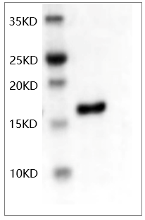 |
| Mouse bFGF protein | PRP1011 | E. coli | > 95 % | Lyophilized powder | 10ug/$48 50ug/$120 100ug/$170 1mg/$900 | 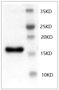 |
| Human IFN-gamma protein, His Tag | PRP1014 | E. coli | > 95 % | Lyophilized powder | 20ug/$48 100ug/$120 500ug/$450 1mg/$800 | 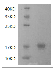 |
| Mouse IFN-gamma protein, His Tag | PRP1015 | E. coli | > 95 % | Lyophilized powder | 20ug/$48 100ug/$120 500ug/$450 1mg/$800 | 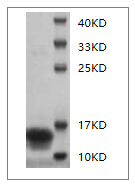 |
Abbkine focuses on the fields of immunology
and cytology, and is committed to innovating and developing various types of
antibodies, proteins, analytical reagents and kits, with a view to becoming a
key promoter in the fields of life science research and development, and drug
development. Here, we present to you the favorite products of protein and
immune research users, from basic immunological products, such as protein
extraction and quantification, to internal reference label antibodies, primary
and secondary antibodies for immunological experiments; cell research users’
favorite Products ranging from dyes and kits for cell state detection,
organelle extraction kits, cell substructure staining and cell metabolism
detection products, to cytokine and protein detection kits for cell culture,
just to help you Research career!
About Abbkine Scientific Co., Ltd.
Abbkine Scientific Co., Ltd. was founded in
2012. Our mission is to become a respected and world-class supplier of
biomedical products and services. Through a clear core strategy and a corporate culture that
promotes learning, innovation, cooperation, and excellence, we will stimulate
our inherent creativity, provide competitive biomedical products and services,
and continue to create maximum value for customers and achieve our mission.
2020年4月1日星期三
How to choose appropriate Catalase (CAT) Activity Assay Kit?
Catalase (CAT) is an antioxidant enzyme commonly found in almost all living organisms. Its main function is to catalyze the decomposition of hydrogen peroxide into water and oxygen and remove hydrogen peroxide in the body, thus preventing cells from being poisoned by H2O2. It is one of the key enzymes in the biological defense system and provides an antioxidant defense mechanism for the body.
In this article, for CAT Activity Assay Kit, the author chose different brands (Cayman Chemical, Abcam, Biovision) and compared them with Abbkine CheKine™ Catalase (CAT) Activity Assay Kit (Cat#: KTB1040) from several aspects such as assay principle, detection, sample type and size/price.
Summary: For CAT Activity Assay Kit, the brand Abbkine and Cayman Chemical are the similar in Assay Principle. Methanol is taken as a reaction substrate. While Abcam and Biovision take H2O2 as a reaction substrate.
But H2O2 is not only a single substrate of catalase (CAT), other peroxidases in the sample may interfere with the experimental results, and the standard curve error may be caused by instability of hydrogen peroxide. While methanol is the unique substrate of catalase, other peroxidases cannot use methanol as substrate, so interference signals are avoided.
Details of different brands of products are as follows,
| Brand | Assay Principle | Sample type | Size/Price |
| Abbkine | This assay kit utilizes the peroxidatic function of catalase for measuring catalase activity, based on the reaction of catalase with methanol, with the presence of an optimal concentration of H2O2. The formaldehyde produced can be measured colorimetrically at OD 540 nm. Therefore, the catalase activity present in the sample is proportional to the signal obtained. | Tissue Homogenate, Cell Lysate, serum, plasma, erythrocyte lysate | 48T/$40 96T/$60 480T/$180 |
| Cayman Chemical | This assay kit utilizes the peroxidatic function of catalase for measuring catalase activity, based on the reaction of catalase with methanol, with the presence of an optimal concentration of H2O2. The formaldehyde produced can be measured colorimetrically at OD 540 nm. Therefore, the catalase activity present in the sample is proportional to the signal obtained. | Tissue Homogenate, Cell Lysate, serum, plasma, erythrocyte lysate | 96T/$270 480T/$490 |
| Abcam | The catalase present in the sample reacts with hydrogen peroxide (H2O2) to produce water and oxygen. The unconverted H2O2 reacts with probe to produce a product that can be measured colorimetrically at OD 570 nm. | Serum, Plasma, Cell Lysate, Tissue Lysate, Urine | 100T/$415 |
| Biovision | The catalase present in the sample reacts with hydrogen peroxide (H2O2) to produce water and oxygen. The unconverted H2O2 reacts with probe to produce a product that can be measured colorimetrically at OD 570 nm. | Tissue Homogenate, Cell Lysate, erythrocyte lysate | 100T/$340 |
About Abbkine Scientific Co., Ltd.
Abbkine serves global scientists in the field of proteomics and cytology and is committed to the innovation and development of various scientific reagents related to proteomics and cytology, expecting to accelerate the pace of life science research and drug discovery. Proteomics products cover the preparation of samples (protein extraction, purification, coupling), protein quantification, antibodies and kits for protein detection. Cytology products involve cytokines (cell culture), cell status detection, cell staining, organelle extraction, cell metabolism and cytopathology reagents (kits). Abbkine relies on the product portfolio and unique marketing support as the main market strategy and product innovation mode, with ultimate aim to facilitate your research career.
2020年3月13日星期五
How to choose appropriate NADP/NADPH Assay Kit?
NADP (coenzyme II) is the coenzyme of many redox reactions, including NADP+ (oxidation type) and NADPH (reduction type). NADP+ is also involved in biosynthesis reactions, such as lipid and nucleic acid synthesis. In animal cells, the oxidation stage of pentose phosphate pathway (PPP) is the main source of NADPH.
In this article, for NADP/NADPH Assay Kit, the author chose different brands (BioAssay Systems, Abcam, Biovision) and compared them with Abbkine CheKine™NADP/NADPH Assay Kit (Cat#: KTB1010) from several aspects such as assay principle, detection, sample type and size/price.
Summary:
These brands are the similar in Assay Principle, Detection and Sample type. Prices are different. The price of Abbkine NADP/NADPH Assay Kit (Cat#: KTB1010) is the lowest.
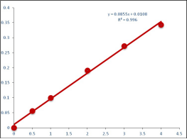
Besides good price, Abbkine NADP/NADPH Assay Kit (Cat#: KTB1010) has other advantages. The detection limit of Abbkine NADP/NADPH Assay Kit is 0.1 µM and linearity up to 4 µM NADP+/NADPH in 96-well plate assay.
Details of different brands for NADP/NADPH Assay Kit are as follows,
| Brand | Assay Principle | Detection | Sample type | Size/Price |
| Abbkine | The assay is based on an enzymatic cycling reaction in which NADP+ is reduced to NADPH. NADPH reacts with a colorimetric probe that produces a colored product which can be measured at 565 nm. | Detects NADP+, NADPH, or total NADP+/NADPH | Tissue Extracts, Cell Lysate | 48T/$90 96T/$150 |
| BioAssay Systems | The assay is based on an enzymatic cycling reaction in which NADP+ is reduced to NADPH. NADPH reacts with a colorimetric probe that produces a colored product which can be measured at 565 nm. | Detects NADP+, NADPH, or total NADP+/NADPH | Tissue Extracts, Cell Lysate | 100T/$379 |
| Abcam | The assay is based on an enzymatic cycling reaction in which NADP+ is reduced to NADPH. NADPH reacts with a colorimetric probe that produces a colored product which can be measured at 450 nm. | Detects NADP+, NADPH, or total NADP+/NADPH | Tissue Extracts, Cell Lysate | 100T/$615 |
| Biovision | The assay is based on an enzymatic cycling reaction in which NADP+ is reduced to NADPH. NADPH reacts with a colorimetric probe that produces a colored product which can be measured at 450 nm. | Detects NADP+, NADPH, or total NADP+/NADPH | Tissue Extracts, Cell Lysate | 100T/$535 |
About Abbkine Scientific Co., Ltd.
Abbkine serves global scientists in the field of proteomics and cytology and is committed to the innovation and development of various scientific reagents related to proteomics and cytology, expecting to accelerate the pace of life science research and drug discovery. Proteomics products cover the preparation of samples (protein extraction, purification, coupling), protein quantification, antibodies and kits for protein detection. Cytology products involve cytokines (cell culture), cell status detection, cell staining, organelle extraction, cell metabolism and cytopathology reagents (kits). Abbkine relies on the product portfolio and unique marketing support as the main market strategy and product innovation mode, with ultimate aim to facilitate your research career.
2020年2月28日星期五
The Statement of Strengthening Epidemic Prevention for COVID-19
Dear Colleagues,
The situation for COVID-19’s outbreak in Wuhan is getting improved in the strong supports of our government and caring people all over the world now. We are very lucky that we all are safe at present, while it’s still very important for us to do well protections. We determine to do below provisional measures during the resumption of production until the end of the outbreak in order to ensure the safe of all colleagues and our clients strictly.
- The establishment of epidemic prevention group, their works contain procurement of protection materials, temperature detection, virus kill, daily patrol and consults ect;
- Purchasing a batch of substance such as medicine alcohol, chlorine disinfectant, medical surgical masks, protective clothing, temperature guns for epidemic prevention;
- The offices, production laboratories, warehouses must be disinfected fully after daily work, and all windows must be opened;
- The temperature detection is needed before and after daily work for everyone, any person has the temperature over 37.3℃ must be sent to the hospital immediately. Early detection, early isolation, early treatment;
- All colleagues must wear mask and the colleagues in production department also must wear protective clothing, gloves, wash and change them frequently;
- The new purchased materials must be kept in isolated room first, and transferred into warehouses or production rooms after being thoroughly sprayed and disinfected with 75% medicine alcohol;
- The high temperature resistant consumables, such as bottle, pipe, experimental spear, must be sterilized via high heat and pressure, and those consumables don’t resistant to high temperature, such as box, bag, must be thoroughly sprayed and disinfected with 75% medicine alcohol to ensure the safe of products during the production;
- During the process of packing box, the inner and outer surface of the box must be sprayed and disinfected with 75% medicine alcohol to ensure the packaging safety;
- Cargos should be transported via reliable express company such as S.F. Express, FedEx, DHL to ensure the transportation safety;
Abbkine Scientific Co.,Ltd.
20th, February 2020
2020年2月20日星期四
How to Select an Antibody&Protein Coupling Kit with Super Cost Performance
1, The common definition of coupling technology
Enzymes, biotin, fluorescent dye, agarose, magnetic beads and other substances which are easy to detect or separate are covalently labeled or conjugated to antibodies, proteins or other small molecular substances by a certain technology.
2, Background of Coupling Technology
Coupling of antibodies, proteins or other small molecules is a necessary step in many types of biological experiments.
For example, in order to detect specific proteins in immunoassays (such as WB/IHC/IF and other immunological experiments), we couple primary antibody or secondary antibody with enzyme, fluorescent dye, etc.
In cell staining (biological imaging) applications, reactive dyes (fluorescent dye) are directly combined with extracellular or intracellular proteins (polypeptides) to produce visible and measurable staining.
Coupling agarose or magnetic beads to proteins or antibodies provides a targeted purification method. For example, the agarose (magnetic beads) coupled antibody used by Abbkine in IP experiments directly saves the use of Protein A/G.
3, Major Disclosure of Coupling Kits of Various Brands (Taking HRP Coupling Kits and Green Fluorescence Coupling Kits as Examples)
| Brand | Sample types | Contents of the kit | Duration of experiment | Size/Price |
| Abbkine | Samples contain free amino group | 1. Activated HRP conjugates solution 2. HRP labeling solution 3. HRP quencher | 3.5-4h | 3*20ug/$100 100ug/$120 3*100ug/$140 1mg/$220 |
| Abcam | Samples contain free amino group | 1. Activated HRP conjugates solution 2. HRP labeling solution 3. HRP quencher | 3.5-4h | 30ug/¥$270 100ug/$275 300ug/$330 1mg/$555 5mg/$1887 |
| ThermoFisher | Samples contain free amino group | 1. Activated HRP conjugates solution 2. HRP labeling solution 3. HRP quencher 4. Protein A/G Column | overnight+>2h | 300µg/$652 |
* How does Abbkine HRP coupling kit remove free HRP (molecular weight 40KD) from binding antibody?
Abbkine HRP coupling kit was designed with this in mind. The kit can provide a low level of free HRP at the end of the reaction. Therefore, no filtration step is required, and the termination solution (quencher) provided in the kit will block the reactive groups of free HRP.
* Protein A/G column in Thermofisher brand is used to remove free HRP.
| Brand | Sample types | Contents of the kit | Duration of experiment | Size/Price |
| Abbkine | Samples contain free amino group | 1. Activated Green fluorescence conjugates solution 2. labeling solution 3. Purification Column | 1.5h | 3*20ug/$120 100ug/$140 3*100ug/$160 1mg/$250 |
| Abcam | Samples contain free amino group | 1. Activated Green fluorescence conjugates solution 2. labeling solution 3. quencher | 0.5-1h | 30ug/$265 100ug/$266 300ug/$415 1mg/$645 |
| ThermoFisher | Samples contain free amino group | 1. Activated Green fluorescence conjugates solution 2. labeling solution 3. Purification Column | 1.5-2h | 100µg/$538 |
* The purpose of the purification column is to separate the unconjugated dye from the coupled product, because the dye is small molecules (molecular weight is about 1KD), and the purification column volume provided by Abbkine is 50KDa, 0.5 ml. The purification column provided by ThermoFisher is also aimed at the purification of antibody (the molecular weight of antibody is about 160KD). If the molecular weight of the protein purified by customers is small or large, they need to equip themselves with appropriate ultrafiltration tubes.
The following recommendations are made for the allocation of ultrafiltration tubes:
| The molecular weight of the sample | Ultrafiltration tubes |
| 6<MW<20K | 3K |
| 20<MW<60K | 10K |
| 60<MW<100K | 30K |
| 100<MW<200K | 50K |
| 200K<MW | 100K |
To sum up, which brand of coupling kit would you choose? As the fans of Abbkine, I'd like to summarize the characteristics and advantages of Abbkine LinKine™ products:
- Optimized Scheme-Follow the standard experimental procedure and obtain the best activity or fluorescence with excellent pre-activated dye, protein ratio and patented solution formula.
- The operation is simple and convenient-the operation can be realized in three steps, and the coupling group is bound to a primary amine site of an antibody or other protein.
- Customization-changing molar ratio, reaction buffer, pH value and other parameters can realize different levels of coupling and activity.
- High Efficiency Purification-The kit includes a purification column to ensure rapid and effective removal of unreacted dyes and excellent protein/antibody recovery.
2020年2月9日星期日
Buy Abbkine selected primary antibodies, get high quality secondary antibodies
Abbkine's selected primary antibodies are the first choice for reliable trust, featuring a selection of popular research targets, strict quality control and quality assurance. We are grateful for everyone's recognition of this product line. In order to allow more customers to use good products and send out high-score articles, we are now launching the activity of buying selected primary antibodies and get high quality secondary antibodies.
Promotion time: From now to December 31th, 2020
Details of activities:
As long as you buy selected primary antibodies, you can get designated high quality secondary antibodies (size: 100ul) for free or at a very preferential price. Designated secondary antibodies are as follows,
| Product name | Cat# | Citation | Size | List Price($) | Preferential Price($) |
| HRP, Goat Anti-Mouse IgG | A21010 | 131 | 100ul | 0 | |
| HRP, Goat Anti-Rabbit IgG | A21020 | 147 | 100ul | 0 | |
| DyLight 488, Goat Anti-Mouse IgG | A23210 | 79 | 100ul | 15 | |
| Dylight 488, Goat Anti-Rabbit IgG | A23220 | 107 | 100ul | 15 | |
| Dylight 549, Goat Anti-Mouse IgG | A23310 | 24 | 100ul | 15 | |
| Dylight 549, Goat Anti-Rabbit IgG | A23320 | 59 | 100ul | 15 | |
| Dylight 594, Goat Anti-Mouse IgG | A23410 | 30 | 100ul | 15 | |
| Dylight 594, Goat Anti-Rabbit IgG | A23420 | 46 | 100ul | 15 | |
| Dylight 649, Goat Anti-Mouse IgG | A23610 | 13 | 100ul | 15 | |
| Dylight 649, Goat Anti-Rabbit IgG | A23620 | 18 | 100ul | 15 | |
| Dylight 800, Goat Anti-Mouse IgG | A23910 | 4 | 100ul | 15 | |
| Dylight 800, Goat Anti-Rabbit IgG | A23920 | 12 | 100ul | 15 | |
| IFKine™ Green Donkey Anti-Mouse IgG | A24211 | 11 | 100ul | 20 | |
| IFKine™ Green Donkey Anti-Rabbit IgG | A24221 | 12 | 100ul | 20 | |
| IFKine™ Red Donkey Anti-Mouse IgG | A24411 | 5 | 100ul | 20 | |
| IFKine™ Red Donkey Anti-Rabbit IgG | A24421 | 22 | 100ul | 20 | |
| IPKine™ HRP, Goat Anti-Mouse IgG LCS | A25012 | 13 | 100ul | 20 | |
| IPKine™ HRP, Mouse Anti-Rabbit IgG LCS | A25022 | 9 | 100ul | 20 | |
| IPKine™ HRP, Goat Anti-Mouse IgG HCS | A25112 | 8 | 100ul | 20 | |
| IPKine™ HRP, Goat Anti-Rabbit IgG HCS | A25222 | 7 | 100ul | 20 |
* IFKine™antibodies: IFKine™ are series of specially optimized secondary antibodies with improved brightness, photostability and less nonspecific hybridization and background. Donkey host and other species of serum/IgG absorbed make it ideal choice for fluorescence staining, especially in fluorescence multiple labelling.
*IPKine™ antibodies: 25 kD or 50 kD protein detection on Western blots after Immunoprecipitation is often suffered from heavy or light chain blotting contamination. Abbkine’s IPKine™ products could solve these problems and bring you satisfying results with good performance.
2020年2月5日星期三
Wuhan Institute of Virology Reveals New Coronavirus (2019-nCoV) may Bat Origin
On February 3, the Wuhan virus research institute of the Chinese academy of sciences/biosafety research center, Wuhan jinyintan hospital and Hubei provincial center for disease prevention and control cooperated in the identification and research of 2019 new coronavirus (2019-nCoV), which caused the Wuhan pneumonia epidemic, and published it online under the topic "a pneumonia outbreak associated with a new coronavirus of disease origin" (the link of the paper is https://www.nature.com/articles/s41586-020-2012-7). Relevant research teams revealed the basic biological characteristics of the coronavirus from nucleic acid detection, serological diagnosis, virus isolation and receptor utilization, providing important clues for epidemic control and drug research and development.
The research team obtained the whole genome sequence of the virus from the early samples of 5 patients. The similarity of the virus sequence from 5 patients reached 99.9%, and the sequence consistency with SARS-CoV was 79.5%. The amino acid analysis of virus conserved protein shows that 2019-nCoV and SARS-CoV belong to SARS-related coronavirus (SARSR-COV). The author further compared 2019-nCoV genome with some gene sequences of coronavirus detected early in the laboratory, and found that the virus is similar to the gene of a coronavirus (TG13 for short) derived from a bat sample. After sequencing the bat sample, the whole genome sequence of bat virus TG13 was obtained, and the sequence consistency of the two viruses was found as high as 96% (Figure 1).
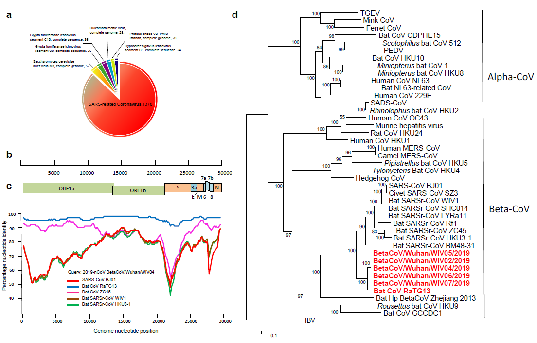
Fig. 1. high-throughput sequencing obtains the whole genome sequence (A) of the virus from a patient sample; Sketch map of 2019-nCoV genome (b); Homology analysis with human SARS virus and bat SARSr-CoV (c); Evolutionary analysis using 2019-nCoV replicase gene (d)
The research team carried out nucleic acid detection on samples of bronchoalveolar lavage fluid (BALF), throat swab (OS) and other samples of patients in different periods, and found that the viral nucleic acid of early samples was about 1000 times higher than that of late samples. By using SARSr-CoV antigen stored in the laboratory at an early stage, they tested the patient's serum samples and found that the patient produced corresponding IgM and IgG antibodies (fig. 2). Subsequently, 2019-nCoV virus was isolated from bronchoalveolar lavage fluid of a critically ill patient, and its intracellularly clear coronavirus particle morphology was observed by electron microscopy (fig. 3).
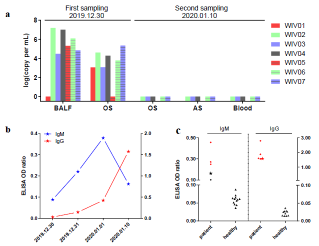
Fig. 2. virus nucleic acid detection in seven patients (a); Serological changes in a patient (B); Detection of Antibody Level in Seven Patients (C)
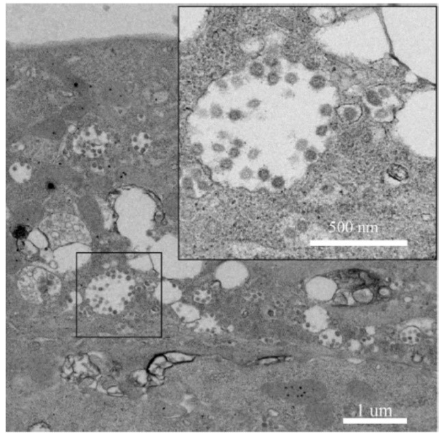
Fig. 3. electron microscopic observation of 2019-ncov in cells
The research team found that 2019-nCoV can infect non-sensitive cells that express human ACE2 (angiotensin converting enzyme, cell receptor of SARS-CoV), indicating that 2019-nCoV can invade cells using receptor of SARS-CoV (fig. 4).
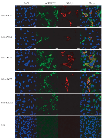
Fig. 4. 2019-nCoV infection in HeLa cells expressing ACE2 from human, bat, pig, civet and mouse
In the race against time and epidemic situation, the study provided more detailed basic biological characteristics of 2019-nCoV virus in a short period of time, and revealed that the natural host of the virus may be bats. The study also showed that 2019-nCoV can infect cells with the receptor of SARS-CoV, laying a working foundation for subsequent epidemic prevention and drug research.
Product Recommendation:
2020年1月15日星期三
Protein Quantification- Reliable and Stable Choice of Excellence
Accurate protein quantification is
necessary for protein-related experiments that involve research in molecular
biology, cell biology, biochemistry, developmental biology, and neuroscience.
Abbkine protein quantification products include Protein Quantification Kits
(BCA Assay and Bradford Assay), SuperLumia ECL HRP Substrate Kit, Protein Gel Flash
Staining Kit, Colorcode Prestained Protein Marker and so on.
Protein
Quantification Kits
Featured products:
1. Protein Quantification Kits (BCA Assay)
• Save time—Easier and 4x faster than
traditional Lowry methods.
• High sensitivity—The detection line is as low as 25ug/ml, and the
minimum detection protein is 0.5ug.
• Good compatibility—unaffected by most ionic and non-ionic
detergents. Compatible with up to 5% SDS, 5% Triton X-100, 5% Tween-20, 60, 80
in the sample.
• Excellent linearity-linear standard curve range: 50– 1000ug/ml.
• Good stability—Protein-to-protein differences are smaller compared
to dye-bound methods.
2. Protein Quantification Kits (Bradford Assay)
• Save time-rapid color development.
• Good compatibility—unaffected by most chemicals. The compatible
concentrations of mercaptoethanol and dithiothreitol in the samples can reach
1mM and 5mM, respectively.
• Good detection range—The detection range is 50-1000ug/ml.
ECL
SuperLumia ECL HRP Substrate Kit developed
by Abbkine is based on the classic peroxidase substrate system innovation and
optimization. It is used to enhance chemiluminescence (ECL) and can directly
replace other expensive ECL products without re-optimizing experimental
conditions. It is the perfect combination of superior quality and affinity
price.
Protein
Gel Flash Staining Kit
Protein staining can be detected in the
range of 10ng to 5μg, which only takes 0.5-1hours. It is about 5-10 times more sensitive than the
traditional Coomassie R-250 dye.

Fig1.
Various volumes of BSA samples were separated on 10% SDS-PAGE, stained with
protein gel flash staining working solution for 30 minutes.
Protein
Marker
Abbkine's Colorcode Prestained Protein Marker (10-180 kDa) contains 3 colors, totaling 10 protein standards (10, 17, 25, 33, 40, 53, 70, 95, 130, 180 kDa). The orange band is 70 kDa; the green band is 10 kDa. Thaw at room temperature. Mix gently and thoroughly to ensure a homogeneous solution.
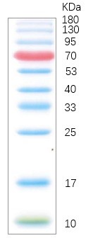
Fig2. Image is from a 15% Tris-glycine gel (SDS-PAGE) transferred to membrane using Abbkine Colorcode Prestained Protein Marker (10-180 kDa).
Abbkine Protein Quantification product
recommendation:
Product name | #Cat | Szie | Price ($) |
Protein Quantification Kit (BCA Assay) | KTD3001 | 500/2000/ 5000T | 45/100/180 |
Protein Quantification Kit (Bradford Assay) | KTD3002 | 500/2000/5000T | 30/70/120 |
Protease Inhibitor Cocktail (100X) | BMP1001 | 1ml/1ml*5 | 40/120 |
SuperLumia ECL HRP Substrate Kit | K22020 | 30/100/200/500ml | 30/75/140/280 |
SuperLumia ECL Plus HRP Substrate Kit | K22030 | 30/100/200/500ml | 45/110/210/440 |
Protein Gel Flash Staining Kit | K21010 | 100/250/500ml | 60/120/200 |
Colorcode Prestained Protein Marker (10-180 kDa) | BMM3001 | 100μl/250μl*2/250μl*20 | 20/62/496 |
Colorcode Prestained Protein Marker (15-130 kDa) | BMM3002 | 100μl/250μl*2/250μl*20 | 16/48/384 |
2020年1月8日星期三
Hard work, Do not forget your initiative mind-—Abbkine Breaks 1400 citations
Since
Abbkine document collection began, as of December 31, 2019, the number of
English articles published by google using Abbkine products has exceeded 1400,
with an impact factor exceeding 5400 points.
Thank you
for your trust and support to Abbkine. We will continuously stimulate our
internal creativity, provide competitive biomedical products and services, and
continuously create maximum value for our customers. With a view to becoming a
respected and world-class provider of biomedical products and services.
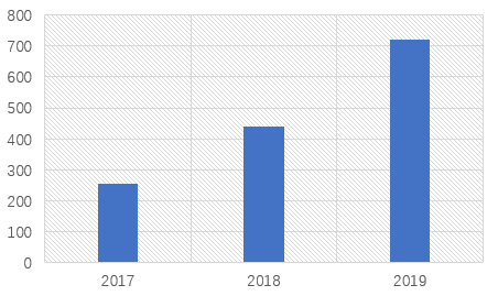
Figure 1:
Number of English Articles Published Using Abbkine Products from 2017 to 2019
In December 2019, Abbkine added 200+ citations. Some high-score citations are as below.
1.LECT2, a Ligand for Tie1, Plays a Crucial Role in Liver Fibrogenesis.
https://doi.org/10.1016/j.cell.2019.07.021
Magazine:
Cell
Impact: 24.38
Abstract:
Liver
fibrosis is a very common condition seen in millions of patients with various
liver diseases, and yet no effective treatments are available owing to poorly
characterized molecular pathogenesis. Here, we show that leukocyte cell-derived
chemotaxin 2 (LECT2) is a functional ligand of Tie1, a poorly characterized
endothelial cell (EC)-specific orphan receptor. Upon binding to Tie1, LECT2
interrupts Tie1/Tie2 heterodimerization, facilitates Tie2/Tie2
homodimerization, activates PPAR signaling, and inhibits the migration and tube
formations of EC. In vivo studies showed that LECT2 overexpression inhibits
portal angiogenesis, promotes sinusoid capillarization, and worsens fibrosis,
whereas these changes were reversed in Lect2-KO mice. Adeno-associated viral vector
serotype 9 (AAV9)-LECT2 small hairpin RNA (shRNA) treatment significantly
attenuates fibrosis. Upregulation of LECT2 is associated with advanced human
liver fibrosis staging. We concluded that targeting LECT2/Tie1 signaling may
represent a potential therapeutic target for liver fibrosis, and serum LECT2
level may be a potential biomarker for the screening and diagnosis of liver
fibrosis.
Products
using from Abbkine:
IPKine™ HRP, Goat Anti-Mouse IgG HCS
(CAT#: A25112)
IPKine™ HRP, Goat Anti-Mouse IgG LCS (CAT#: A25012)
2. EMS1 and BRI1 control separate biological processes via extracellular domain diversity and intracellular domain conservation.
https://www.nature.com/articles/s41467-019-12112-w
Magazine: Nature
Communications volume
Impact: 12.19
Abstract: In
flowering plants, EMS1 (Excess Microsporocytes 1) perceives TPD1 (Tapetum
Determinant 1) to specify tapeta, the last somatic cell layer nurturing pollen
development. However, the signaling components downstream of EMS1 are
relatively unknown. Here, we use a molecular complementation approach to
investigate the downstream components in EMS1 signaling. We show that the EMS1
intracellular domain is functionally interchangeable with that of the
brassinosteroid receptor BRI1 (Brassinosteroid Insensitive 1). Furthermore,
expressing EMS1 together with TPD1 in the BRI1 expression domain could
partially rescue bri1 phenotypes, and led to the dephosphorylation of BES1, a
hallmark of active BRI1 signaling. Conversely, expressing BRI1 in the EMS1
expression domain could partially rescue ems1 phenotypes. We further show that
PpEMS1 and PpTPD1 from the early land plant Physcomitrella patens could
completely rescue ems1 and tpd1 phenotypes, respectively. We propose that EMS1 and
BRI1 have evolved distinct extracellular domains to control different
biological processes but can act via a common intracellular signaling pathway.
Products using from Abbkine:
Anti-Plant Actin Mouse Monoclonal Antibody (3T3) (CAT#: A01050)
3. PLK4 deubiquitination by Spata2‐CYLD suppresses NEK7‐mediated NLRP3 inflammasome activation at the centrosome.
https://www.embopress.org/doi/abs/10.15252/embj.2019102201
Magazine: EMBO
JOURNAL
Impact: 10.55
Abstract:
The innate
immune sensor NLRP3 assembles an inflammasome complex with NEK7 and ASC to
activate caspase‐1 and drive the maturation of proinflammatory cytokines IL‐1β
and IL‐18. NLRP3 inflammasome activity must be tightly controlled, as its
over‐activation is involved in the pathogenesis of inflammatory diseases. Here,
we show that NLRP3 inflammasome activation is suppressed by a centrosomal
protein Spata2. Spata2 deficiency enhances NLRP3 inflammasome activity both in
the macrophages and in an animal model of peritonitis. Mechanistically, Spata2
recruits the deubiquitinase CYLD to the centrosome for deubiquitination of
polo‐like kinase 4 (PLK4), the master regulator of centrosome duplication.
Deubiquitination of PLK4 facilitates its binding to and phosphorylation of NEK7
at Ser204. NEK7 phosphorylation in turn attenuates NEK7 and NLRP3 interaction,
which is required for NLRP3 inflammasome activation. Pharmacological or
shRNA‐mediated inhibition of PLK4, or mutation of the NEK7 Ser204
phosphorylation site, augments NEK7 interaction with NLRP3 and causes increased
NLRP3 inflammasome activation. Our study unravels a novel centrosomal
regulatory pathway of inflammasome activation and may provide new therapeutic
targets for the treatment of NLRP3‐associated inflammatory diseases.
Products using from Abbkine:
IFKine™ Green Donkey Anti-Mouse IgG (CAT#: A24211)
4. Cross-Microbial Protection via Priming a Conserved Immune Co-Receptor through Juxtamembrane Phosphorylation in Plants
https://doi.org/10.1016/j.chom.2019.10.010
Magazine: Cell
Host & Microbe
Impact: 10.5
Abstract: Living
organisms can be primed for potentiated responses to recurring stresses based
on prior experience. However, the molecular basis of immune priming remains
elusive in plants that lack adaptive immunity. Here, we report that bacterial
challenges can prepare plants for fungal attacks by inducing juxtamembrane
phosphorylation of CERK1, the co-receptor indispensable for signaling in
response to the fungal elicitor chitin. This phosphorylation is mediated by
BAK1, a co-receptor for signaling in response to multiple elicitors. BAK1
interacts with CERK1, and loss of BAK1 reduces priming phosphorylation of
CERK1. Juxtamembrane phosphomimetic mutations of CERK1 confer accelerated
chitin responses and fortified fungal resistance without triggering
constitutive immunity, whereas juxtamembrane phosphodeficient mutations
diminish bacteria-induced protection against fungal infection. These findings
reveal that crosstalk between cell-surface immune co-receptors can prime
defense and demonstrate that juxtamembrane phosphorylation of plant
receptor-like kinases can occur independent of kinase activation to place the
protein into a prime state.
Products using from Abbkine:
Anti-GST Tag Mouse Monoclonal Antibody (2A8) (CAT#: A02030)
5. Extracellular vesicles of carcinoma-associated fibroblasts creates a pre-metastatic niche in the lung through activating fibroblasts
https://molecular-cancer.biomedcentral.com/articles/10.1186/s12943-019-1101-4
Magazine: Molecular
Cancer
Impact: 9.17
Abstract: Carcinoma-associated
fibroblasts (CAFs) have been known to promote cancer progression by modifying
the primary tumor microenvironment. We aimed to elucidate the intercellular
communication between CAFs and secondary organs in salivary adenoid cystic
carcinoma (SACC) metastasis.
Products using from Abbkine:
FITC, Goat Anti-Rabbit IgG (CAT#: A22120)
Dylight 488, Goat Anti-Rabbit IgG
(CAT#: A23220)
Dylight 549, Goat Anti-Rabbit IgG (CAT#: A23320)
Please learn more details from https://www.abbkine.com/publications/ .
2020年1月3日星期五
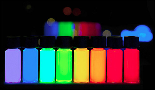
The third generation of fluorescent dye AbFluor™/highly water-soluble fluorescent dye
AbFluor™ dyes are a
series of highly water-soluble fluorescent dyes introduced by Abbkine after
many years of research, covering the ultraviolet, visible and near-infrared
spectrum, suitable for labeling biological macromolecules, especially proteins
and nucleic acids. Their excellent hydrophilicity enables protein-conjugated
labeling to be performed easily in aqueous media, while minimizing the need for
organic solvents. AbFluor™ dyes not only have better fluorescence performance
than other fluorescent dyes (such as FITC, TRITC, Alexa Fluor and Dylight
dyes), but also the coupling products are more stable.
Why recommend
AbFluor™ fluorescent dyes?
With innovative
modifications to the core structure of the dye, AbFluor™ dyes have more
brilliant properties than other commercial dyes, including stronger fluorescent
brightness, better light stability, wider pH tolerance range, and better
penetration and lower background. As a third-generation fluorescent dye,
AbFluor™ dyes not only perform better than traditional dyes (such as FITC,
TRITC, and Cy dyes), but also perform better than other second-generation
commercial dyes (such as Alexa Fluor, Dylight and IRDye).
1. AbFluor™ dyes
have excellent core structure, stable reactive groups, and good light
stability, ensuring high stability and high labeling efficiency of
bioconjugation.
2. High water
solubility (>100 mg/ml), which minimizes fluorescence quenching; it is also
easily soluble in other polar solvents, such as DMSO, DMF, methanol and
ethanol.
3. A wide range of
fluorescence spectrum, covering ultraviolet, visible, and near-infrared, to
meet most of the experimental needs of researchers.
4. Good pH
tolerance, stable in pH 2-11.
Taking AbFluor™ 488
as an example, the following is an IF comparison chart of different dye effects:
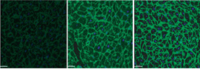
Different dye : FITC Alexa Fluor 488 AbFluor™ 488
So, are you excited,
and what are you waiting for???
Abbkine AbFluor™ product recommendation:
Product name | #Cat | Szie | Price ($) |
LinKine™ AbFluor™ 405 Labeling Kit | KTL0510 | 3*20μg/100μg/ 3*100μg/1mg | 1200/1400/ 1600/2500 |
LinKine™ AbFluor™ 488 Labeling Kit | KTL0520 | 3*20μg/100μg/ 3*100μg/1mg | 1200/1400/ 1600/2500 |
LinKine™ AbFluor™ 555 Labeling Kit | KTL0530 | 3*20μg/100μg/ 3*100μg/1mg | 1200/1400/ 1600/2500 |
LinKine™ AbFluor™ 594 Labeling Kit | KTL0540 | 3*20μg/100μg/ 3*100μg/1mg | 1200/1400/ 1600/2500 |
LinKine™ AbFluor™ 647 Labeling Kit | KTL0560 | 3*20μg/100μg/ 3*100μg/1mg | 1200/1400/ 1600/2500 |
LinKine™ AbFluor™ 680 Labeling Kit | KTL0580 | 3*20μg/100μg/ 3*100μg/1mg | 1200/1400/ 1600/2500 |
LinKine™ AbFluor™ 770 Labeling Kit | KTL0590 | 3*20μg/100μg/ 3*100μg/1mg | 1200/1400/ 1600/2500 |